McGill Case 321: Difference between revisions
Jump to navigation
Jump to search
(Created page with "{{McGillcase| |previouspage= McGill Case 320 |previousname= McGill Case 320 |nextpage= McGill Case 322 |nextname= McGill Case 322 }} [[File:E321.jpg|thumb|600px|left|This is ...") |
(No difference)
|
Latest revision as of 23:04, 19 February 2012
|

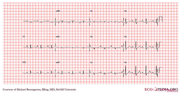
This is an electrocardiogram from a young woman with Ebstein's anomaly. In this condition the right atrium is enlarged with downward a displacement of the tricuspid valve into the right ventricle. This partially explains the tall peaked p waves in the inferior leads. Note the P wave in lead II is greater than 2 mm in height. There is also a first-degree heart block which is often seen with these patients. Curiously higher grade blocks are rare. The QRS complex shows a right axis deviation and a terminal S wave in the V 6 derivation. Atypical right bundle branch block can be seen with this condition.