McGill Case 87: Difference between revisions
Jump to navigation
Jump to search
Created page with "{{McGillcase| |previouspage= McGill Case 86 |previousname= McGill Case 86 |nextpage= McGill Case 88 |nextname= McGill Case 88 }} [[File:E0007871.jpg|thumb|600px|left|These ar..." |
No edit summary |
||
| (3 intermediate revisions by the same user not shown) | |||
| Line 6: | Line 6: | ||
}} | }} | ||
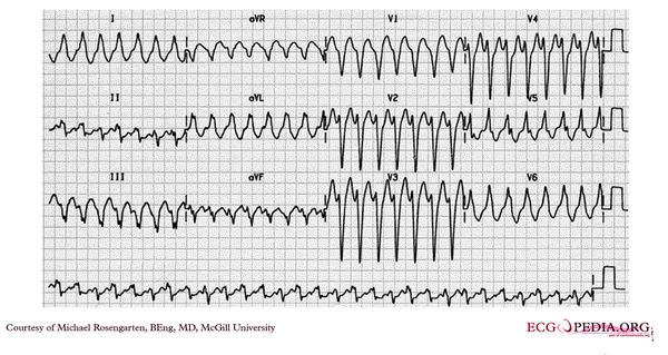
[[File: | [[File:E000789.jpg|thumb|600px|left|This is an electrocardiogram from an elderly woman with palpitations. | ||
The | The cardiogram shows a wide complex tachycardia with a left bundle branch morphology at a rate of about 160/min. The R wave in V2 is broad and the time from the beginning of the QRS in V2 to the peak of the S wave is longer than 80 ms. No P wave activity is clearly seen. The cardiogram suggests ventricular tachycardia. The patient has done well since this cardiogram on flecainide and metoprolol.]] | ||
The | |||
Latest revision as of 04:45, 15 February 2012

|
