McGill Case 89: Difference between revisions

From ECGpedia
Jump to navigation Jump to search
(Created page with "{{McGillcase| |previouspage= McGill Case 88 |previousname= McGill Case 88 |nextpage= McGill Case 90 |nextname= McGill Case 90 }} [[File:E000789.jpg|thumb|600px|left|This is a...")
 
No edit summary
 
Line 6: Line 6:
}}
}}


[[File:E000789.jpg|thumb|600px|left|This is an electrocardiogram from an elderly woman with palpitations.
[[File:E0007891.jpg|thumb|600px|left|70 year old patient, previous myocardial infarction, on a beta-blocker, presents with the rhythm below. No response to adenosine or to verapamil IV.]]
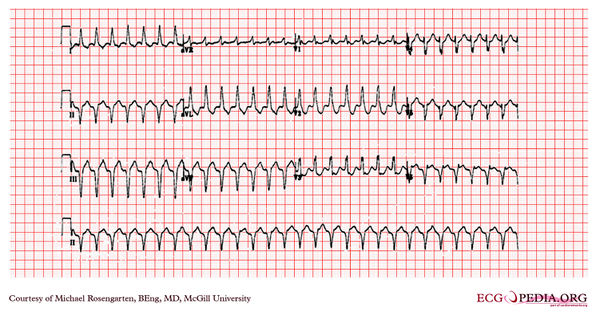
The cardiogram shows a wide complex tachycardia with a left bundle branch morphology at a rate of about 160/min. The R wave in V2 is broad and the time from the beginning of the QRS in V2 to the peak of the S wave is longer than 80 ms. No P wave activity is clearly seen. The cardiogram suggests ventricular tachycardia. The patient has done well since this cardiogram on flecainide and metoprolol.]]
 
[[File:E0007892.jpg|thumb|600px|left|After cardioversion.]]

Latest revision as of 04:33, 15 February 2012

McGill logo.png
This case report is kindly provided by Michael Rosengarten from McGill and is part of the McGill Cases. These cases come from the McGill EKG World Encyclopedia.


Previous Case: McGill Case 88 | Next Case: McGill Case 90

70 year old patient, previous myocardial infarction, on a beta-blocker, presents with the rhythm below. No response to adenosine or to verapamil IV.
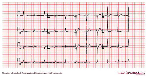
After cardioversion.