McGill Case 67: Difference between revisions
Jump to navigation
Jump to search
No edit summary |
No edit summary |
||
| Line 11: | Line 11: | ||
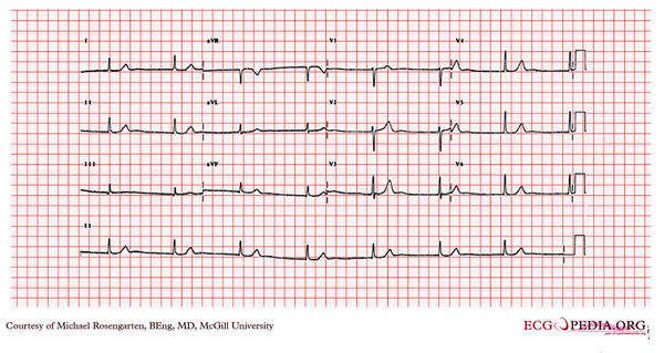
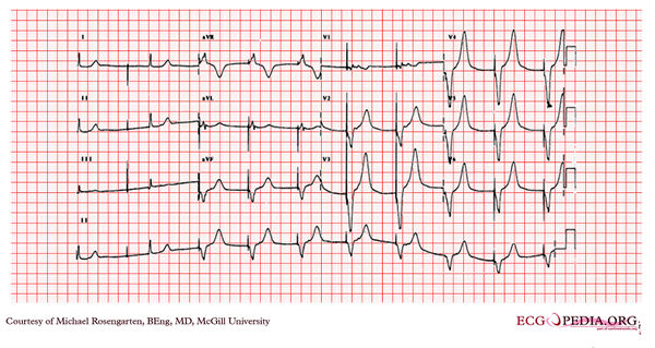
[[File:E0007682.jpg|thumb|600px|left|This tracing was taken in the intensive care unit after a temporary pacing wire (soft semi-floater) was placed via the right internal jugular vein. The lead paced the ventricle well, but the patient immediately complained of moderate chest pain, better with sitting up.]] | [[File:E0007682.jpg|thumb|600px|left|This tracing was taken in the intensive care unit after a temporary pacing wire (soft semi-floater) was placed via the right internal jugular vein. The lead paced the ventricle well, but the patient immediately complained of moderate chest pain, better with sitting up.]] | ||
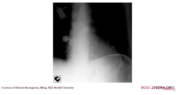
[[File: | [[File:E0007683_68.jpg|thumb|600px|left|This was an X-ray taken the day after the insertion of the temporary pacing lead. The patient continued to have chest pain.]] | ||
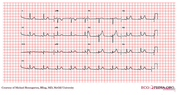
[[File:E0007684.jpg|thumb|600px|left|This cardiogram was taken at the peak of the chest pain.]] | [[File:E0007684.jpg|thumb|600px|left|This cardiogram was taken at the peak of the chest pain.]] | ||
Revision as of 01:47, 15 February 2012
| This case report is kindly provided by Michael Rosengarten from McGill and is part of the McGill Cases. These cases come from the McGill EKG World Encyclopedia.
|


