Basics: Difference between revisions
| Line 31: | Line 31: | ||
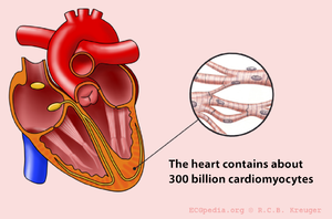
[[Image:Hart_cells_en.png|thumb|The heart consists of approximately 300 trillion cells]] | [[Image:Hart_cells_en.png|thumb|The heart consists of approximately 300 trillion cells]] | ||
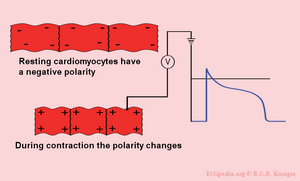
[[Image:cells_in_rest_en.png|thumb|In rest the heart cells are negatively charged. Trough the depolarization by surrounding cells they become positively charged and they contract.]] | [[Image:cells_in_rest_en.png|thumb|In rest the heart cells are negatively charged. Trough the depolarization by surrounding cells they become positively charged and they contract.]] | ||
[[Image: | [[Image:Ion_currents_en.jpg|thumb|During the depolarization sodium-ions stream inwards the cell. Subsequently the calcium-ions stream inwards the cell. These calcium-ions give the actual muscular contraction. Finally the potassium-ions stream out of the cell. During the repolarisation the ion concentration is corrected. On the ECG an action potential wave coming towards is shown as a positive result. Here the ECG electrode is represented as an eye.]] | ||
The individual [[action potential|action potentials]] of the individual cardiomyocytes are averaged. The final result which is shown on the ECG is actually the average of trillions of microscopic electronical signals. | The individual [[action potential|action potentials]] of the individual cardiomyocytes are averaged. The final result which is shown on the ECG is actually the average of trillions of microscopic electronical signals. | ||
{{clr}} | {{clr}} | ||
Revision as of 22:25, 10 December 2006
Introduction
The aim of this course is to understand and recognize the normal ECG and to be able to interprete abnormalities. The course is divided in different sections. First the basics will be presented. This is followed by the interpretation of the normal ECG. Then abnormalities are discussed: (ischemia, arrhythmias and Miscellaneous). Finally the real world is presented in practice ECGs.
The American College of Cardiology has published a list of abnormalities a professional should be able to recognise. It is advisable to go through this list at the end of this course in order to recognise areas that need your attention.
How do I begin reading an ECG?
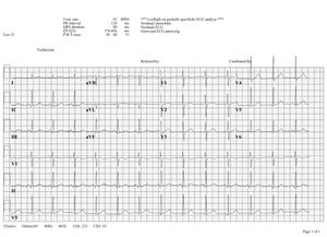
Click on the ECG to see an enlargement. Where do we look at watching an ECG?
- top left are the patient's information, name, sex and date of birth
- at the right of that are below each other the heart frequency, the conduction time intervals (PQ,QRS,QT/QTc), and the cardiac axis (P-top axis, QRS axis and T-top axis)
- farther to the right is the interpretation of the ECG written (this often misses in a 'fresh' ECG, but later the interpretation of the cardiologist or computer will be added)
- down left is the 'paper speed' (25mm/s on the horizontal ax), the sensitivity (10mm/mV) and the filter's frequency (40Hz, filters noise from eg. lights)
- finally there is a calibration on the ECG, on the beginning of every lead is a vertical block that shows how high 1mV is. So the height and depth of these signals are a measurement for the voltage. If this is not the set 10mm, there is something wrong with the machine.
- further we have the ECG leads themselves of course, what these are will be discussed below.
Note the lay-out of the paper is for every machine diffrent, but all of the information above can be found somewhere mostly.
What does the ECG register?
An ECG is a registration of the heart's electric activity.
Just like skeletal muscles, the heart is electrically stimulated to contract. This stimulation is also called activation or excitation. Cardiac muscles are electrically charged in rest. The surface of the cell is positively charged relative to the inside (resting potential). If the cardiac muscle cells are electrically stimulated (depolarisation: the outside of the depolarized region is positively charged relative to the inside) and there comes an action potential, the cells contract. Because of the spreading of the impulse through the heart, the electric field changes continually in size and direction. The ECG is a graphical visualisation of the electric signals in the heart.
The ECG is a sum of the action potentials from millions of cardiomyocytes
The individual action potentials of the individual cardiomyocytes are averaged. The final result which is shown on the ECG is actually the average of trillions of microscopic electronical signals.
The electric discharge of the heart
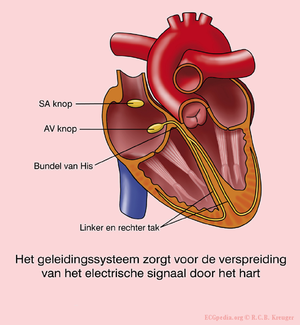
In the sinal node (SA node) are pacemakercells which determine the heart frequency.
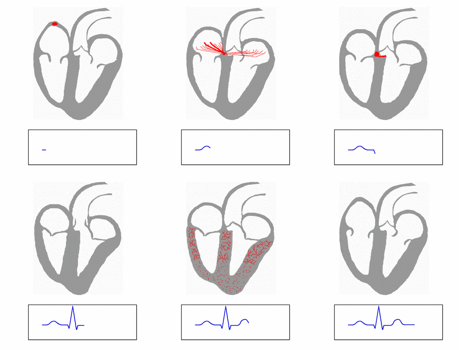
First the atria depolarise and contract, after that the ventricles
The electrical signal between the atria and the ventricles goes from the sinus node, via the atria to the AV-node (atrioventricular overgang) to the His bundle and subsequently to the right and left bundle branche, which uitmonden in a fijnvertakt network of Purkinje fibers.
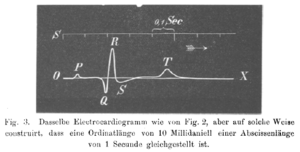
The different ECG waves
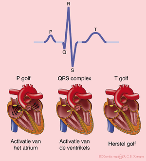
De P top onstaat door depolarisatie van de atria. Deze golf begint in de SA-knoop, waarna er geleiding plaatsvind naar de rechter en vervolgens naar de linker atria. Repoolarisatie van de atria wordt normaal gesproken niet waargenomen op een ECG. De repolarisatie valt samen met het QRS-complex en is van een kleine omvang (minder weefsel dan de ventrikels).
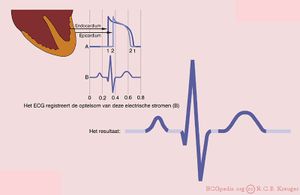
Het QRS complex is een middeling van de depolarisatiegolven van de endomyocardiale (binnenste) en epicardiale (buitenste) spiercellen. Of te wel de depolarisatie van de venrtrikels. Doordat de endomyocardiale cellen net iets eerder epolariseren dan de epicardiale spiercellen, ontstaat het typische QRS patroon.
De T golf ontstaat door repolarisatie van de ventrikelcellen. Tijdens de T golf is er geen spieractiviteit (het hart staat stil).
Éen hartslag omvat een boezemsystole (contractie atria --> p-top), kamersystole (kamer contractie --> ORS-complex) en de rustfase (T-top) tussen twee slagen.
Zie ook deze [animatie van de hartcyclus]
De oorsprong van de U golf is onbekend. Mogelijk duidt deze golf op "afterdepolarisaties" van de ventrikels.
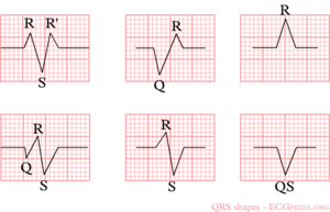
De letters QRS worden op verschillende manieren geschreven om verschillende vormen aan te duiden:
- Q: eerste negatieve deflectie na de p-top. Als die er niet is, is er dus geen Q
- R: positieve deflectie
- S: negatieve deflectei na de R-top
- met kleine letters (q, r, s) worden kleine deflecties aangegeven. Bijvoorbeeld: qRS = kleine q, hoge R, diepe S.
- R` (uitspraak: r-accent): wordt gebruikt om een tweede R-top aan te geven (bijvoorbeeld bij een rechter bundeltakblok)
Zie ook enkele voorbeelden op de afdeling rechts.
The history of the ECG
De geschiedenis van het ECG gaat ver terug.
In 1843 beschreef Emil Du bois-Reymond een Duitse fydioloog voor het eerste de "actiepotentiaal" van een spiercontractie. Hij maakte gebruik van een gevoelige galvanometer voor zijn metingen. Hierin zat een spoel met 5 km draad verwerkt. Du Bios Reymond benoemde de verschillende golven: "o" was het stabiele equilibrium en hij was de eerste die de letters p, q, r en s gebruikte. Du Bois-Reymond, E. Untersuchungen uber thierische Elektricitat. Reimer, Berlin: 1848.
In 1850 beschreef M. Hoffa hoe hij onregelmatige contracties van de ventrikels veroorzaakte door hondenharten een electrische schok te geven. Hoffa M, Ludwig C. 1850. Einige neue versuche uber herzbewegung. Zeitschrift Rationelle Medizin, 9: 107-144
In 1887 publiceerde de Engelse fysioloog Augustus D. Waller uit Londen het eerste menselijke electrocardiogram. Hij gebruikte een capillair-electrometer. Waller AD. A demonstration on man of electromotive changes accompanying the heart's beat. J Physiol (London) 1887;8:229-234
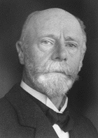
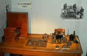
Willem Einthoven (1860-1927) introduceerde in 1893 de term 'electrocardiogram'. Hij beschreef in 1895 hoe hij een galvanometer gebruikte om de electrische activiteit van het hart op te tekenen. In 1924 heeft hij hiervoor de Nobelprijs gekregen als grondlegger van het huidige ECG. Hij sloot zijn electrodes aan op de patient en liet het electrische verschil tussen twee electrodes uitschrijven door een galvanometer. Wij spreken nog steeds van de afleidingen van Einthoven. De snaar galvanometer (zie Image) werd in zijn tijd geroemd als het eerste instrument dat een klinische implicatie had.
In 1905 neemt Einthoven het eerste 'telecardiogram' op vanuit het ziekenhuis naar zijn laboratorium 1,5 km verderop.
In 1906 publiceert Einthoven het eerste artikel waarin een serie (afwijkende) ECG bevindingen worden beschreven: linker en rechter ventrikelhypertrofie, linker en rechter atriumdilatatie, de U golf, notching van het QRS comples, ventriculaire extrasystolen, bigemini, boezemflutter en totaal AV blok. Einthoven W. Le telecardiogramme. Arch Int de Physiol 1906;4:132-164
The ECG electrodes
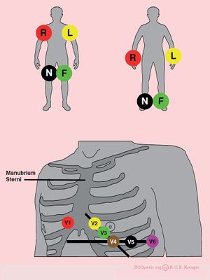
Electric activity that goes through the heart, can be measured by external (skin)electrodes. The electrocardiogram (ECG) registers these activities from these electrodes which have been attached on diffrent places on the body. In total, twelve leads are to be calculated using ten electrodes.
The ten electrodes are:
- the extremity leads:
- LA - left arm
- RA - right arm
- N - neutral, on the right leg (= electrisch aarde of nulpunt ten opzichte waarvan de electrische spanning wordt gemeten)
- F - foot, on the left leg
It makes no diffrence whether the electrodes will be attached proximal or distal on the extremities. Echter, it is better not to afwisselen it. (eg. an electrode on the left shoulder and one on the right wrist).
- the chest leads:
- V1 - geplaatst in de 4e intercostaalruimte rechts van het borstbeen
- V2 - geplaatst in de 4e intercostaalruimte links van het borstbeen
- V3 - geplaatst halverwege tussen V2 en V4
- V4 - geplaatst in de 5e intercostaalruimte in de tepellijn
- V5 - geplaatst halverwege tussen V4 en V6
- V6 - geplaatst in de axillairlijn op dezelfde hoogte als V4
Met behulp van deze 10 electrodes kunnen dus 12 afleidingen uitgeschreven worden. Er zijn 6 extremiteitsafleidingen en 6 voorwandsafleidingen.
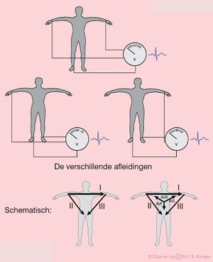
The extremity leads
De extremiteitsafleidingen zijn:
- I van rechter naar linker arm
- II van rechter arm naar linker been
- III van linker arm naar linker been
Een makkelijk te onthouden ezelsbruggetje: Afleiding I + Afleiding III = Afleiding II Hierbij wordt gebruik gemaakt van de hoogten en/of diepten, onafhankelijk van de golf (QRS, P of T). Voorbeeld: is in Afleiding I het QRS complex 3mm hoog, in Afleiding III 9mm, dan zal de hoogte van het QRS-complex in afleiding II 12mm bedragen.
Daarnaast zijn er electrisch afgeleide afleidingen. Deze hebben als centrum het electrisch gemiddelde van de extremiteitsafleidingen (ongeveer het hart zelf dus).
- AVL wijst naar de Linker arm
- AVR naar de Rechter arm
- AVF naar de voeten (Feet)
De letter a staat voor "augmented" (versterkt) en de letter V voor "voltage".
(aVR + aVL + aVF = 0)
The chest leads
De voorwandsafleidingen (V1,V2,V3,V4,V5 en V6) 'kijken' vanuit hun borstelectrodes naar het electrisch gemiddelde. Dus in feite naar het centrum van het hart.
Voorbeeld: V1 zit vlakbij de rechter kamer en het rechter atrium en signalen vanuit die gebieden geven in deze afleiding de grootste uitslag. V6 zit vlakbij de laterale (=zijkant) van de linker hartkamer, hier worden signalen vanuit de linker hartkamer het best geregistreerd.
Special leads
Bij een onderwandinfarct worden soms extra afleidingen gebruikt:
- Bij een zogenaamd rechts uitgepoold ECG behouden V1 en V2 hun plaats. V3 tm V6 worden op dezelfde plaats gezet, maar dan langs de rechterkant van het borstbeen. Op het ECG moet aangegeven worden dat het om een Rechts-ECG gaat. V4R (V4 maar dan rechts uitgepoold) is een gevoelige afleiding om een rechterkamerinfarct te diagnostiseren.
- Afleidingen V7-V8-V9 worden gebruikt om een posteriorinfarct aan te tonen. Hierbij wordt doorgepoold ter hoogte van V6 naar de rug. Een posteriorinfarct is meestal ook goed te zien in V2 (maar dan 'op de kop', zie ook het hoofdstuk ischemie, dus deze afleidingen worden zelden gebruikt.
Technische problemen met het ECG
Draadverwisselingen
Het komt nogal eens voor dat één van de afleidingsdraden niet goed aangesloten wordt. Het is handig om dit te kunnen herkennen, want anders kan je verkeerde conclusies trekken.
Denk bij een 'vreemd' ECG daarom aan een dradenverwisseling. Een van de meest voorkomende fouten is het verwisselen van de linker en rechter arm. Dit uit zich in een negatieve afleiding in I. Dit zou ook door een rechter asdraai kunnen komen, maar dat is heel zeldzaam.
Veel voorkomende verwisselingen zijn, omkering van:
- rechter en linker arm electroden;
- omkering van afleiding II en III
- omkering van de afleingen aVR en aVL
- linker arm en linker been:
- omkering van afleiding I en II
- omkering van afleiding aVF en aVF
- inversie van sfleiding III
- rechter arm en linker been:
- inversie va afleiding I, II en III
- omkering van afleidingen aVR en aVF
Men kan draadverwisseling en dextrocardie van elkaar onderscheiden door eveneens naar de precordiale afleidingen te kijken. Dextrocardia toont R-golf inversie i.t.t. omkering van de electroden.
Storing
- Bewegingsartefacten
- Tremor
- Electrische storing
- Verkeerde filter-instelling

