McGill Case 38: Difference between revisions
Jump to navigation
Jump to search
Created page with "{{McGillcase| |previouspage= McGill Case |previousname= McGill Case |nextpage= McGill Case |nextname= McGill Case }} [[File:E000623.jpg|thumb|600px|left|From a Holter rec..." |
No edit summary |
||
| (2 intermediate revisions by the same user not shown) | |||
| Line 1: | Line 1: | ||
{{McGillcase| | {{McGillcase| | ||
|previouspage= McGill Case | |previouspage= McGill Case 37 | ||
|previousname= McGill Case | |previousname= McGill Case 37 | ||
|nextpage= McGill Case | |nextpage= McGill Case 39 | ||
|nextname= McGill Case | |nextname= McGill Case 39 | ||
}} | }} | ||
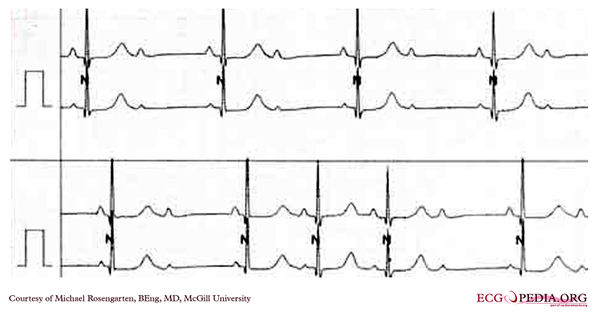
[[File: | [[File:E000738.jpg|thumb|600px|left|These two strips from the same patient show a 2:1 block on the top tracing and a Mobitz II A/V block on the lower one. Note that with 2:1 block you can not tell if this is a Mobitz I or II. Mobitz II is seen below as the PR does not change before and after the non-conducted P wave.]] | ||
Latest revision as of 03:10, 11 February 2012

|
