McGill Case 39: Difference between revisions
Jump to navigation
Jump to search
No edit summary |
No edit summary |
||
| (2 intermediate revisions by the same user not shown) | |||
| Line 6: | Line 6: | ||
}} | }} | ||
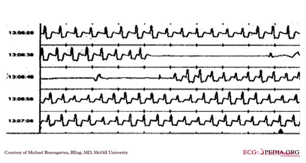
[[File: | [[File:E0007391.jpg|thumb|600px|left|The first panel is a compressed view of the recording. Note the black triangle at the bottom of the last strip. This is where the patient froze the memory of the loop recording and was just after having a near syncopal episode. | ||
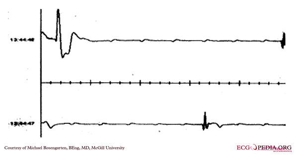
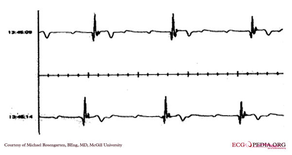
The strips below are expanded to about normal speed and are from the same recording but at a later time when the patient was again symptomatic. Here we clearly see complete heart block with sinus p waves moving through the recordings.]] | |||
[[File:E0007392.jpg|thumb|600px|left|The first panel is a compressed view of the recording. Note the black triangle at the bottom of the last strip. This is where the patient froze the memory of the loop recording and was just after having a near syncopal episode. | |||
The strips below are expanded to about normal speed and are from the same recording but at a later time when the patient was again symptomatic. Here we clearly see complete heart block with sinus p waves moving through the recordings.]] | |||
[[File:E0007393.jpg|thumb|600px|left|The first panel is a compressed view of the recording. Note the black triangle at the bottom of the last strip. This is where the patient froze the memory of the loop recording and was just after having a near syncopal episode. | |||
The strips below are expanded to about normal speed and are from the same recording but at a later time when the patient was again symptomatic. Here we clearly see complete heart block with sinus p waves moving through the recordings.]] | |||
Latest revision as of 03:04, 11 February 2012

|


