Sinus Node Rhythms and Arrhythmias: Difference between revisions
No edit summary |
mNo edit summary |
||
| Line 1: | Line 1: | ||
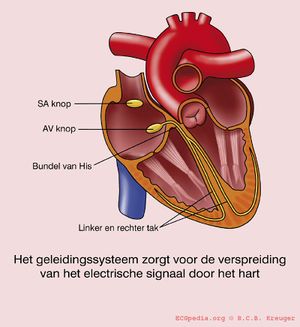
[[Image:geleidingssysteem.jpg|thumb|The conducting system handels the spreading of an electrical signal through the heart. The normal sinus rhythm begins in the sinus node and goes via the AV node to the His bundle where it splits via the right and left bundle branch.]] | [[Image:geleidingssysteem.jpg|thumb|The conducting system handels the spreading of an electrical signal through the heart. The normal sinus rhythm begins in the sinus node and goes via the AV node to the His bundle where it splits via the right and left bundle branch.]] | ||
This part is about the normal ECG. The normal heart rhythm is | This part is about the normal ECG. The normal heart rhythm is sinus rhythm. That means that the rhythm has its origin in the sinal node, the heart's fastest physiological impulse generator. | ||
The sinus node (SA) is located in the upper part of the wall of the right atrium. When the sinus node generates an electrical impulse, first the cells of the right atrium depolarise, then the cells of the left atrium, the AV (atrioventricular) node follows and at last the ventricles are stimulated via the His bundle. | The sinus node (SA) is located in the upper part of the wall of the right atrium. When the sinus node generates an electrical impulse, first the cells of the right atrium depolarise, then the cells of the left atrium, the AV (atrioventricular) node follows and at last the ventricles are stimulated via the His bundle. | ||
With this knowledge it is quite simple to recognise sinus rhythm on the ECG. | With this knowledge it is quite simple to recognise normal sinus rhythm on the ECG. | ||
{{clr}} | {{clr}} | ||
| Line 10: | Line 10: | ||
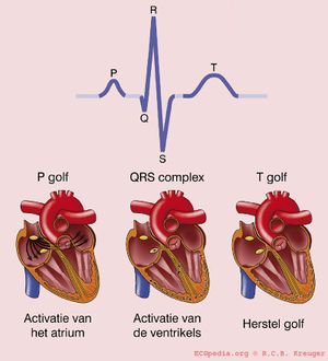
[[Image:QRSverklaring.jpg|thumb| During a normal sinus rhythm, after every atrial contraction (P-top) follows a ventricular contraction (QRS complex).]] | [[Image:QRSverklaring.jpg|thumb| During a normal sinus rhythm, after every atrial contraction (P-top) follows a ventricular contraction (QRS complex).]] | ||
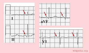
[[Image:normalSR.jpg|thumb|Normal sinus rhythm with a positive P-top in I, II and AVF, and a biphasic P-top in V1.]] | [[Image:normalSR.jpg|thumb|Normal sinus rhythm with a positive P-top in I, II and AVF, and a biphasic P-top in V1.]] | ||
*A P-top (atrial contraction) | *A P-top (atrial contraction) ''gaat vooraf aan'' the QRS complex | ||
*Every P follows a QRS complex | *Every P follows a QRS complex | ||
*The rhythm is regular, but varies slightly while breathing | *The rhythm is regular, but varies slightly while breathing | ||
Revision as of 22:05, 13 October 2006
This part is about the normal ECG. The normal heart rhythm is sinus rhythm. That means that the rhythm has its origin in the sinal node, the heart's fastest physiological impulse generator.
The sinus node (SA) is located in the upper part of the wall of the right atrium. When the sinus node generates an electrical impulse, first the cells of the right atrium depolarise, then the cells of the left atrium, the AV (atrioventricular) node follows and at last the ventricles are stimulated via the His bundle.
With this knowledge it is quite simple to recognise normal sinus rhythm on the ECG.
The properties of normal sinus rhythm (see also Basics):
- A P-top (atrial contraction) gaat vooraf aan the QRS complex
- Every P follows a QRS complex
- The rhythm is regular, but varies slightly while breathing
- The frequency is between 60 and 100 beats per minute
- The P-top's maximum height is 2.5mm in II and/or III
- The P-top is positive in I and II, and biphasic in V1
These last two definitions will be discussed in the topic P-top morphology.
Heart rhythms which are no sinus rhythm will be discussed in the topic rhythm disorders.


