McGill Case 24: Difference between revisions
Jump to navigation
Jump to search
Created page with "{{McGillcase| |previouspage= McGill Case 23 |previousname= McGill Case 23 |nextpage= McGill Case 25 |nextname= McGill Case 25 }} [[File:E000607.jpg|thumb|600px|left|A patient..." |
No edit summary |
||
| (3 intermediate revisions by 2 users not shown) | |||
| Line 6: | Line 6: | ||
}} | }} | ||
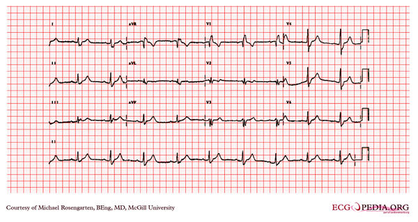
[[File: | [[File:E000724.jpg|thumb|600px|left|This is an electrocardiogram from an elderly woman who had previously undergone surgery for recurrent ventricular tachycardia. She was being treated with Tambacor and metoprolol. | ||
The cardiogram shows sinus rhythm with a wide QRS of 159ms consistent with a RBBB and a rightward axis suggesting right posterior hemi-block. The PR interval is slightly prolonged at 2121ms. The poor R wave progression seen best in lead V2 suggests previous anterior wall MI.]] | |||
Latest revision as of 05:24, 10 February 2012

|
