Puzzle 2009 04 Answer: Difference between revisions
m Created page with '{{NHJ| |mainauthor= '''A.A.M. Wilde''' |edition= 2009:04,172 }} thumb A 71-year-old female patient presents to your outpatients clinic with an irregula…' |
mNo edit summary |
||
| (One intermediate revision by one other user not shown) | |||
| Line 12: | Line 12: | ||
==Answer== | ==Answer== | ||
Indeed, the heart rhythm is irregular. The P wave morphology is abnormal with negative P waves in the inferior leads, an almost negative P wave in lead I and a positive P wave in lead aVR. This indicates a lower left atrial origin. The cycle length of this rhythm is 640 | |||
ms (rate slightly lower than 100 beats/min). The wide intervals on the ECG result from a blocked atrial impulse every fourth beat. The block is at the level of the origin of the atrial impulse, i.e. an exit block of the focal impulse. Atrioventricular and ventricular conduction are normal. Repolarisation is abnormal and might be explained by the use of digoxin. Atrial tachycardia with AV block might be seen in the setting of toxic digoxin concentrations but in this case the level of block is not at the atrioventricular junction. So, there is no reason to consider this cause. | |||
Electrical cardioversion could bring this abnormal rhythm back to normal sinus rhythm. | |||
Latest revision as of 19:04, 3 August 2011
| Author(s) | A.A.M. Wilde | |
| NHJ edition: | 2009:04,172 | |
| These Rhythm Puzzles have been published in the Netherlands Heart Journal and are reproduced here under the prevailing creative commons license with permission from the publisher, Bohn Stafleu Van Loghum. | ||
| The ECG can be enlarged twice by clicking on the image and it's first enlargement | ||

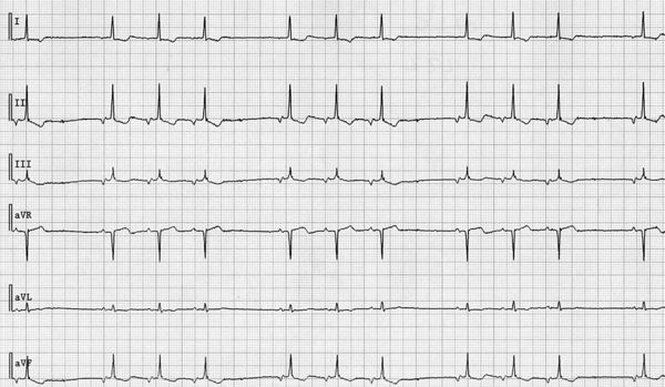
A 71-year-old female patient presents to your outpatients clinic with an irregular heart rhythm. The complaints started a few weeks ago and seem to worsen. Her cardiac history reveals the use of digoxin prescribed by her general physician for an irregular, much faster, heart beat a few years ago. The present symptoms are different from those. Physical examination did not reveal any abnormalities except the irregular heart rhythm. Additional cardiological examinations (X-ray, echo) were without abnormalities. Part of the ECG is shown in figure 1. Only the extremity leads are shown (standard calibration).
What is your diagnosis?
Answer
Indeed, the heart rhythm is irregular. The P wave morphology is abnormal with negative P waves in the inferior leads, an almost negative P wave in lead I and a positive P wave in lead aVR. This indicates a lower left atrial origin. The cycle length of this rhythm is 640 ms (rate slightly lower than 100 beats/min). The wide intervals on the ECG result from a blocked atrial impulse every fourth beat. The block is at the level of the origin of the atrial impulse, i.e. an exit block of the focal impulse. Atrioventricular and ventricular conduction are normal. Repolarisation is abnormal and might be explained by the use of digoxin. Atrial tachycardia with AV block might be seen in the setting of toxic digoxin concentrations but in this case the level of block is not at the atrioventricular junction. So, there is no reason to consider this cause. Electrical cardioversion could bring this abnormal rhythm back to normal sinus rhythm.