P Wave Morphology
| «Step 4:Heart axis | Step 6: QRS morphology» |
The P wave morphology can reveal right or left atrial stretch or atrial arrhythmias and is best determined in leads II and V1 during sinus rhythm.
| |||||||||


| Characteristics of a normal p wave:[1] |
|---|
|
Elevation or depression of the PTa segment (the part between the p wave and the beginning of the QRS complex) can result from Atrial infarction or pericarditis.
If the p-wave is enlarged, the atria are enlarged.
If the P wave is inverted, it is most likely an ectopic atrial rhythm not originating from the sinus node.
Examples
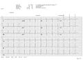
An example of normal sinus rhythm.
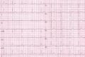
Another example of normal sinus rhythm.

