Answer MI 18
| This page is part of Cases and Examples |
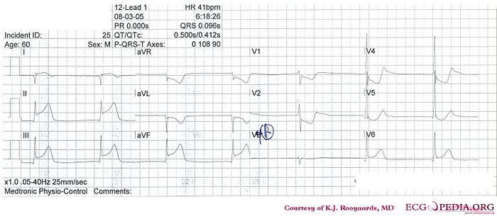
Where is this myocardial infarction located?
Answer
- Following the 7+2 steps:
- Rhythm
- Regular rhythm with narrow QRS complexes without P waves. Probably a nodal escape rhythm with either atrial standstill or atrial fibrillation with AV block
- Heart rate
- 41 bpm
- Conduction (PQ,QRS,QT)
- PQ: not applicable QRS: 100ms QT: 450ms QTc: 370ms
- Heartaxis
- Negative in I, positive in II and AVF, thus a right axis deviation.
- P wave morphology
- No P waves present.
- QRS morphology
- Narrow QRS, no pathologic Q waves, normal precordial R wave progression. A notch is seen in the terminal part of the QRS complex in V5/V6.
- ST morphology
- ST elevation in I, II, III, AVF, V6. ST depression in AVR, AVL, V1-V5. No ST deviation in V4R
- Compare with the old ECG (not available, so skip this step)
- Conclusion?
- Rhythm
Inferior-posterior-lateral myocardial infarction with a nodal escape rhythm - probably due to RCX occlusion
