P Wave Morphology
liacc4tchi
| «Step 4:Heart axis | Step 6: QRS morphology» |
| Author(s) | J.S.S.G. de Jong, MD | |
| Moderator | J.S.S.G. de Jong, MD | |
| Supervisor | ||
| some notes about authorship | ||
The p wave morphology can reveal right or left atrial stretch.
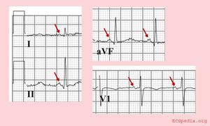
The P-wave morphology is best determined in leads II and V1 during sinus rhythm.
The normal P wave
Characteristics of a normal p wave:[1]
- The maximal height of the P wave is 2.5 mm in leads II and / or III
- The p wave is positive in II and AVF, and bifasic in V1
- The p wave duration is usually shorter than 0.12 seconds
Elevation or depression of the PTa segment (the part between the p wave and the beginning of the QRS complex) can result from Atrial infarction or pericarditis.
If the p-wave is enlarged, the atria are enlarged.
