Repolarization (ST-T,U) Abnormalities
| Author(s) | I.A.C. van der Bilt | |
| Moderator | I.A.C. van der Bilt | |
| Supervisor | ||
| some notes about authorship | ||
Repolarization can be influenced by many factors, including electrolyte shifts, ischemia, structural heart disease (cardiomyopathy) and (recent) arrhythmias. Although T/U wave abnormalities are rarely specific for one disease, it can be useful to know which conditions can change repolarization.
- Early repolarization (normal variant)
- Juvenile T waves (normal variant)
- Nonspecific abnormality, ST segment and/or T wave
- ST and/or T wave suggests ischemia
- ST suggests injury
- ST suggests ventricular aneurysm
- Q-T interval prolonged
- Prominent U waves
- Cardiac Memory|Cardiac Memory
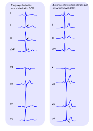
Early repolarization is a normal variant of the ST segment, seen in 2-5% of patients, especially young men. Early repolarization is characterized by elevation of the J point and the beginning of the ST segment as well as elevation of the ST segment itself[1]. The ST segment may be concave up (cup-like) or concave (dome-like). These findings are most often present in the middle chest leads V2-V5. Recently a different form of early repolarization has been associated with idiopathic ventricular fibrillation. This form is most often seen in lead II and consists of a 'hump' in the tail of the QRS complex, without ST elevation.
References
- Wellens HJ. Early repolarization revisited. N Engl J Med. 2008 May 8;358(19):2063-5. DOI:10.1056/NEJMe0801060 |
- Tikkanen JT, Anttonen O, Junttila MJ, Aro AL, Kerola T, Rissanen HA, Reunanen A, and Huikuri HV. Long-term outcome associated with early repolarization on electrocardiography. N Engl J Med. 2009 Dec 24;361(26):2529-37. DOI:10.1056/NEJMoa0907589 |
