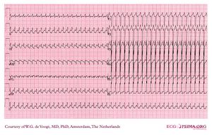
Example 25
Jump to navigation
Jump to search
- Following the 7+2 steps:
- Rhythm
- The ECG shows a regular fast rhythm. P waves are not easily discernible but might well be located just after each QRS complex (for example in lead II)
- Heart rate
- almost 300bpm
- Conduction (PQ,QRS,QT)
- The PQ and QT interval is hard to define, QRS complexes are small
- Heartaxis
- QRS positive in I and AVF: normal heart axis
- P wave morphology
- Negative P-waves in inferior leads
- QRS morphology
- Normal QRS complexes, normal axis, normal R-wave progression in precordial leads (V1 to V6)
- ST morphology
- Because of the fast rhythm the ST segments and T waves are not easy to interpret, there seems to be ST depression with negative T waves in the inferior and lateral leads .
- Compare with the old ECG (not available, so skip this step)
- Conclusion?
- Rhythm
A regular small-QRS tachycardia at about 300bpm with normal looking QRS complexes is most likely an Atrial flutter with 1:1 conduction over the AV node. Carotid sinus massage or adenosine would slow down AV conduction and would uncover the typical 'saw-tooth' P-waves at 300bpm.
It might be that such a fast ventricular rhythm causes myocardial ischaemia without coronary artery disease. Most likely this is a young individual, older individuals often do not have AV node-conduction abilities at these high rates and they would show for example 1:2 or 1:3 AV-block whith the atrial frequency still at 300bpm but with the ventricles activated at 150 or 100bpm.
