Wide Complexes Intervening Regular Sinus Rhythm - 2: Difference between revisions

From ECGpedia
Jump to navigation Jump to search
mNo edit summary
mNo edit summary
Line 18: Line 18:
[[Puzzle_2007_1_33_Answer|Answer]]
[[Puzzle_2007_1_33_Answer|Answer]]
[[Image:Puzzle_2007_1_33_fig2.png|Figure 2|thumb]]
[[Image:Puzzle_2007_1_33_fig2.png|Figure 2|thumb]]
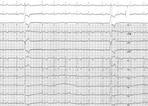
The ECG shows sinus rhythm with a mean rate of 60
beats/min. There is some sinus arrhythmia present.
Conduction intervals are normal (PR 150 ms, QRS
90 ms). Repolarisation abnormalities are present in the
right precordial leads V1 to V3 (terminal negative T
waves) and the ST-T segment in lead V4 is flat.
Two wide complexes are seen, the third and tenth
QRS complex. Both share a similar morphology with
a left bundle branch block and an almost vertical axis.
There is no discernible P wave prior to these wide QRS
complexes and the first is followed by a complete compensatory
pause in contrast to the second which does
not influence the underlying sinus rhythm. The
morphology of the wide complexes, the lack of P waves
and the presence of a complete compensatory pause all
point to a ventricular origin of this activity. Based on
the morphology, an origin in the right ventricular
outflow track (RVOT) is likely. In combination with
the ST-T segment abnormalities one should consider
arrhythmogenic right ventricular dysplasia (ARVD/C)
and additional imaging should be performed.
There are several electrocardiographic peculiarities
in this electrocardiogram. First, the second ventricular
extrasystole is not intervening with the sinus rhythm.
This is referred to as an interpolated ventricular extrasystole.
This is a relatively rare phenomenon usually
seen with a slow heart rate. In this case the heart rate
is not really slow.
Second, the conducting P wave of the P wave
following this extrasystole has a longer PR interval
(figure 2). This phenomenon is referred to as concealed
retrograde conduction; i.e. the ventricular extrasystole
conducts retrogradely in the AV node slowing the
anterograde conducting P wave. In the case of the first
extrasystole retrograde conduction actually blocks the
antegrade P wave completely.
In conclusion, this is a sinus rhythm with ventricular
extrasystoles originating in the RVOT (one of them
interpolated) with retrograde penetrance in the AV
node. The ST-T segment abnormalities in combination
with the extrasystole’s origin suggest the presence of
ARVC.

Revision as of 19:31, 8 October 2007

Author(s) A.A.M. Wilde
NHJ edition: 2007:01,033
These Rhythm Puzzles have been published in the Netherlands Heart Journal and are reproduced here under the prevailing creative commons license with permission from the publisher, Bohn Stafleu Van Loghum.
The ECG can be enlarged twice by clicking on the image and it's first enlargement
Figure 1

A 38-year-old male patient presents with palpitations. He is not suffering from syncope or dizziness and has no other complaints. The family history bears no peculiarities. Physical examination of the heart reveals an irregular heart beat. Auscultation is normal and there are no signs of left or right heart failure. The ECG is depicted in figure 1.

What is your diagnosis and should there be any further investigation?
Answer

Figure 2