Ventricular Fibrillation: Difference between revisions
Jump to navigation
Jump to search
mNo edit summary |
m (→Examples) |
||
| Line 16: | Line 16: | ||
==Examples== | ==Examples== | ||
<gallery> | <gallery> | ||
Image:Rhythm_ventricular_fibrillation.png|Ventricular Fibrillation (VF or V-fib) | |||
Image:ECG_SR_to_VF_in_INF_MI.jpg|VF develops in a patient with an [[inferior MI]] | Image:ECG_SR_to_VF_in_INF_MI.jpg|VF develops in a patient with an [[inferior MI]] | ||
Image:ECG_SR_to_VF_.jpg|Seven sinus beats are follow by a ventricular extrasystole (with R on T phenomenon), resulting in ventricular fibrillation | Image:ECG_SR_to_VF_.jpg|Seven sinus beats are follow by a ventricular extrasystole (with R on T phenomenon), resulting in ventricular fibrillation | ||
</gallery> | </gallery> | ||
Revision as of 13:53, 23 July 2007
| This is part of: Ventricular Arrhythmias |
| {{{locatieafbeelding}}} | |
| Atrial rate | 60-100 bpm |
| Ventricular rate | 400-600 bpm |
| Regularity | irregular |
| Origin | ventricles |
| P-wave | AV-dissociation |
| Effect of adenosine | none |
| Example ECG: {{{example}}} | |
| Example ECG2: {{{example2}}} | |
Ventricular fibrillation (VF or V-fib) is chaotic depolarisation of the ventricles. Mechanically this results in an arrested cardiac pump function and immediate death. VF can only be treated by immediate defibrillation. If you consider ventricular fibrillation in a conscious patient, than you should look for a technical problem with the ECG, eg. movement or electrical interference.
Examples
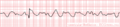
Ventricular Fibrillation (VF or V-fib)
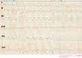
VF develops in a patient with an inferior MI
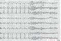
Seven sinus beats are follow by a ventricular extrasystole (with R on T phenomenon), resulting in ventricular fibrillation

