Search results
Jump to navigation
Jump to search
Page title matches
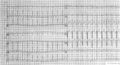
File:De-ECG Aflutt.jpg (800 × 435 (108 KB)) - 08:20, 15 May 2012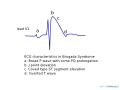
File:Brugada ecg characteristics.svg (1,000 × 750 (36 KB)) - 05:55, 22 May 2013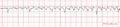
File:De-ECG Parkinson.png (799 × 178 (42 KB)) - 11:38, 10 May 2012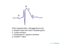
File:De-Brugada ecg characteristics.png (800 × 600 (41 KB)) - 16:11, 10 May 2012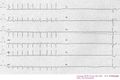
File:De-SSS ecg 001.jpg (800 × 536 (68 KB)) - 07:20, 15 May 2012
File:De-ECG Aflutt 1to1.jpg (800 × 260 (58 KB)) - 08:22, 15 May 2012
File:De-ECG lead angulation.png (800 × 600 (56 KB)) - 23:45, 18 May 2012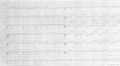
File:De-ECG VWI 2wk.jpg (800 × 439 (75 KB)) - 10:52, 24 May 2012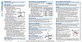
File:ECG reference card thumbnail.jpg (954 × 492 (146 KB)) - 04:51, 24 April 2009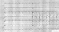
File:De-ECG RBTB LAtrD.jpg (800 × 435 (103 KB)) - 07:34, 10 May 2012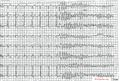
File:De-ECG SR to VF .jpg (800 × 543 (178 KB)) - 22:28, 18 May 2012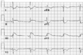
File:De-ECG atrial rhythm now sr.png (800 × 536 (127 KB)) - 09:08, 26 May 2012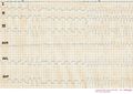
File:De-ECG SR to VF in INF MI.jpg (800 × 565 (109 KB)) - 22:28, 18 May 2012


Page text matches
File:Torsades de Pointes TdP.png 12-lead ECG of Torsades de Pointes (TdP) in a 56-year-old white female with a potassium(765 × 429 (135 KB)) - 01:15, 4 June 2012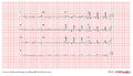
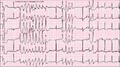
File:E295.jpg The ECG is in sinus rhythm and the QRS is markedly widened with a QRS duration of 2 When presented as a puzzler the correct interpretation of this ECG was not received, only suggestions of ventricular bigemini where given.(3,292 × 1,887 (5.78 MB)) - 08:06, 21 February 2012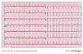
File:DVA2467.jpg |Description = ICD ECG E_A(3,000 × 1,942 (3.24 MB)) - 11:10, 8 March 2011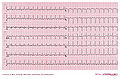
File:DVA2583.jpg |Description = ICD ECG E_A(3,000 × 1,969 (3.3 MB)) - 11:59, 9 March 2011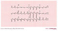
File:E000799.jpg |Description = This is an ECG from a 70 year old woman with severe coronary artery disease and recurrent(3,004 × 1,599 (4.51 MB)) - 01:27, 15 February 2012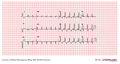
File:E000722.jpg |Description = An ECG from a cardiac transplant patient showing sinus rhythm with one PVC, left a(3,004 × 1,599 (4.49 MB)) - 22:30, 10 February 2012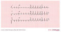
File:E000777.jpg |Description = An ECG from a cardiac transplant patient showing sinus rhythm with one PVC, left a(3,004 × 1,599 (4.49 MB)) - 02:16, 15 February 2012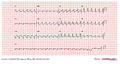
File:E0007921.jpg |Description = This is an ECG from a 67 year old man with a low ejection fraction. The patient is followe(3,004 × 1,599 (4.44 MB)) - 04:15, 15 February 2012
File:E0003163.jpg |Description = This is an ECG strip and an audio recording from a patient checking his Medtronic VVI pace(3,004 × 512 (1.06 MB)) - 00:41, 19 February 2012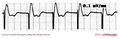
File:E0003180.jpg ...ker one month before. The pacer was programed to VVI mode and AAI mode and ECG recordings were made. Also a PA and lateral chest x-ray was taken. The ques(3,004 × 985 (730 KB)) - 01:30, 19 February 2012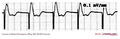
File:E301.jpg ...ker one month before. The pacer was programed to VVI mode and AAI mode and ECG recordings were made. Also a PA and lateral chest x-ray was taken. The ques(3,004 × 968 (718 KB)) - 08:16, 21 February 2012
File:E315.jpg 2 ventricular premature beats are also shown in this ECG(3,292 × 1,887 (5.85 MB)) - 09:57, 21 February 2012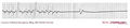
File:E000763.jpg ...g. The 12 lead EKG showed only left atrial abnormality. A surface averaged ECG was considered abnormal with a QRSD of 112ms, a RMS40 of 19.8 ms and a LAS(3,004 × 649 (711 KB)) - 00:54, 15 February 2012
File:E0007082.jpg ...piratory rates. There was no obvious swelling of his lower extremities. An ECG revealed a RBBB not present on previous ECGs. A CXR revealed no failure or(2,644 × 1,599 (3.41 MB)) - 07:22, 10 February 2012
File:E0007081.jpg ...piratory rates. There was no obvious swelling of his lower extremities. An ECG revealed a RBBB not present on previous ECGs. A CXR revealed no failure or(2,644 × 1,599 (3.41 MB)) - 07:17, 10 February 2012


