Sinus Node Rhythms and Arrhythmias: Difference between revisions
No edit summary |
mNo edit summary |
||
| (82 intermediate revisions by 12 users not shown) | |||
| Line 1: | Line 1: | ||
[[ | {{nav| | ||
|previouspage=Basics | |||
|previousname=Basics | |||
|nextpage=Rate | |||
|nextname=Step 2: Heart Rate | |||
}} | |||
{{authors| | |||
|mainauthor= [[user:Drj|J.S.S.G. de Jong]] | |||
|moderator= [[user: Vdbilt|I.A.C. van der Bilt]] | |||
|supervisor= | |||
}} | |||
Please read the [[Introduction|introduction to the 7+2 step plan]] first. | |||
The sinus node (SA) is located in the | The sinus node (SA) is located in the roof of the right atrium. It is the fastest physiological pacemaker. When the sinus node generates an electrical impulse, the surrounding cells of the right atrium depolarize. Then the cells of the left atrium, the AV (atrioventricular)node, follow, and at last the ventricles are stimulated via the His bundle. | ||
{{#widget:Html5media | |||
|url=https://en.ecgpedia.org/images/a/a3/Normal_SR.mp4 | |||
|width=640 | |||
|height=360 | |||
}} | |||
{ | With this knowledge it is quite simple to recognize normal sinus rhythm on the ECG. | ||
{| class="wikitable" | |||
[[ | !Criteria for normal sinus rhythm (see also [[Basics]]): | ||
|- | |||
* | | | ||
*A [[P wave morphology]] P wave (atrial contraction) precedes every QRS complex | |||
* | *The rhythm is regular, but varies slightly during respirations | ||
* | *The rate ranges between 60 and 100 beats per minute | ||
* | *The P waves maximum height at 2.5 mm in II and/or III | ||
*The P wave is positive in I and II, and biphasic in V1 | |||
|} | |||
As you can see, knowledge of [[Rate|heart rate]] and [[P wave morphology]] are necessary to determine the rhythm. We have put Rhythm as step 1 as it is of great importance. Arrhythmias include the most life-threatening ECG abnormalities. In most settings, however, the rhythm will be sinus. | |||
If the rhythm is not sinus, the '''[[Arrhythmias|Arrhythmias algorithm]]''' should be followed. | |||
==Sinus arrhythmias== | |||
Some variants of sinus rhythm exist: | |||
*[[Asystole]] | |||
*[[Sinustachycardia|Sinustachycardia (>100 beats per minute)]] | |||
*[[Sinusbradycardia|Sinusbradycardia (<60 beats per minute)]] | |||
*[[Sinusarrest|Sinus arrest or pause]] | |||
*[[Sino-atrial_exit_block|Sino-atrial exit block]] | |||
*[[Sick Sinus Syndrome]] | |||
*[[Sinus Arrhythmia]] | |||
Arrhythmias are discussed in the [[Arrhythmias]] chapter. | |||
If the heart rate exceeds 100 bpm, the [[Arrhythmias#Tachyarrhythmias|tachcyardia flow chart]] should be followed. | |||
{{clr}} | |||
==Examples== | |||
<gallery> | |||
Image:Normaal ecg.jpg|An example of normal sinus rhythm. | |||
Image:Nsr.jpg|Another example of normal sinus rhythm. | |||
</gallery> | |||
{{clr}} | {{clr}} | ||
[[nl:Ritme]] | [[nl:Ritme]] | ||
Latest revision as of 21:10, 14 January 2021
| «Basics | Step 2: Heart Rate» |
| Author(s) | J.S.S.G. de Jong | |
| Moderator | I.A.C. van der Bilt | |
| Supervisor | ||
| some notes about authorship | ||
Please read the introduction to the 7+2 step plan first.
The sinus node (SA) is located in the roof of the right atrium. It is the fastest physiological pacemaker. When the sinus node generates an electrical impulse, the surrounding cells of the right atrium depolarize. Then the cells of the left atrium, the AV (atrioventricular)node, follow, and at last the ventricles are stimulated via the His bundle.
With this knowledge it is quite simple to recognize normal sinus rhythm on the ECG.
| Criteria for normal sinus rhythm (see also Basics): |
|---|
|
As you can see, knowledge of heart rate and P wave morphology are necessary to determine the rhythm. We have put Rhythm as step 1 as it is of great importance. Arrhythmias include the most life-threatening ECG abnormalities. In most settings, however, the rhythm will be sinus.
If the rhythm is not sinus, the Arrhythmias algorithm should be followed.
Sinus arrhythmias
Some variants of sinus rhythm exist:
- Asystole
- Sinustachycardia (>100 beats per minute)
- Sinusbradycardia (<60 beats per minute)
- Sinus arrest or pause
- Sino-atrial exit block
- Sick Sinus Syndrome
- Sinus Arrhythmia
Arrhythmias are discussed in the Arrhythmias chapter.
If the heart rate exceeds 100 bpm, the tachcyardia flow chart should be followed.
Examples
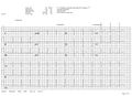
An example of normal sinus rhythm.
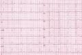
Another example of normal sinus rhythm.

