ST Morphology: Difference between revisions
m (→ST elevatie) |
mNo edit summary |
||
| Line 1: | Line 1: | ||
{{ActiveDiscuss}} | {{ActiveDiscuss}}{{authors| | ||
|mainauthor= [[user:Drj|J.S.S.G. de Jong, MD]] | |||
|advisor= | |||
|coauthor= | |||
|moderator= [[user:Drj|J.S.S.G. de Jong, MD]] | |||
|editor= | |||
}} | |||
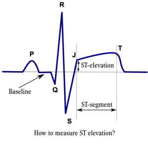
The ''ST segment'' represents the ventricular repolarisation. Repolarisation follows upon contraction and depolarisation. During repolarisation the cardiomyocytes elongate and prepare for the next heartbeat. This process takes much more time than the depolarisation. Repolarisation is not passive elongation by stretch, it is an active process during which energy is consumed. On the ECG, the repolarisation fase starts at the junction, or ''j point'', and continues until the ''T wave''. The ST segment is normally at or near the baseline. | |||
The ''T wave'' is usually concordant with the QRS complex. Thus if the QRS complex is positive in a certain lead (the area under the curve above the baseline is greater than the area under the curve below the baseline) than the T wave usually is positive too in that lead. Accordingly the T wave is normally upright or positive in leads I, II, AVL, AVF and V3-V6. The T wave is negative in V1 and AVR. The T wave flips around V2, but there is some genetical influence in this as in Blacks the T wave usually flips around V3. | |||
The T wave angle is the result of small differences in the duration of the repolarisation between the endocardial and epicardial layers of the left ventricle. The endocardial myocytes need a little more time to repolarise (about 22 msec). This difference causes an electrical current from the endocardium to the epicardium, which reads as a positive signal on the ECG.<cite>braunwald</cite> | |||
==ST elevation== | |||
==ST | |||
[[Image:stelevatie_en.png|thumb|ST elevatie wordt gemeten 1,5 of 2mm (=60ms of 80ms) na het j-punt.<cite>Gibbons</cite>]] | [[Image:stelevatie_en.png|thumb|ST elevatie wordt gemeten 1,5 of 2mm (=60ms of 80ms) na het j-punt.<cite>Gibbons</cite>]] | ||
De belangrijkste oorzaak van '''ST elevatie''' is '''acute [[Ischemia]]'''. Andere oorzaken zijn <cite>Wang</cite><cite>Werf</cite>: | De belangrijkste oorzaak van '''ST elevatie''' is '''acute [[Ischemia]]'''. Andere oorzaken zijn <cite>Wang</cite><cite>Werf</cite>: | ||
| Line 59: | Line 62: | ||
#Wang pmid=14645641 | #Wang pmid=14645641 | ||
#Werf pmid=12559937 | #Werf pmid=12559937 | ||
#brauwnald isbn=0808923056 | |||
</biblio> | </biblio> | ||
<analytics uacct="UA-807577-6"></analytics> | <analytics uacct="UA-807577-6"></analytics> | ||
Revision as of 14:26, 20 May 2007
Some statements may be disputed, incorrect or biased. |
| Author(s) | J.S.S.G. de Jong, MD | |
| Moderator | J.S.S.G. de Jong, MD | |
| Supervisor | ||
| some notes about authorship | ||
The ST segment represents the ventricular repolarisation. Repolarisation follows upon contraction and depolarisation. During repolarisation the cardiomyocytes elongate and prepare for the next heartbeat. This process takes much more time than the depolarisation. Repolarisation is not passive elongation by stretch, it is an active process during which energy is consumed. On the ECG, the repolarisation fase starts at the junction, or j point, and continues until the T wave. The ST segment is normally at or near the baseline.
The T wave is usually concordant with the QRS complex. Thus if the QRS complex is positive in a certain lead (the area under the curve above the baseline is greater than the area under the curve below the baseline) than the T wave usually is positive too in that lead. Accordingly the T wave is normally upright or positive in leads I, II, AVL, AVF and V3-V6. The T wave is negative in V1 and AVR. The T wave flips around V2, but there is some genetical influence in this as in Blacks the T wave usually flips around V3.
The T wave angle is the result of small differences in the duration of the repolarisation between the endocardial and epicardial layers of the left ventricle. The endocardial myocytes need a little more time to repolarise (about 22 msec). This difference causes an electrical current from the endocardium to the epicardium, which reads as a positive signal on the ECG.[1]
ST elevation

De belangrijkste oorzaak van ST elevatie is acute Ischemia. Andere oorzaken zijn [3][4]:
- Acute pericarditis: alle afleidingen geëleveerd behalve aVR
- Longembolie : V1 en aVR
- Hypothermie : V3-V6, II, III en aVF
- Hypertrofische cardiomyopathie : V3-V5 (kan ook nog V6)
- Hyperkaliëmie: V1-V2 (V3)
- Acute neurologische aandoeningen: alle afleidingen, met name V1-V6
- Acute sympathische stress: alle afleidingen, met name V1-V6
- Brugada syndroom.
- Contusio Cordis.
- Aneurysma Cordis
ST depressie
De belangrijkste oorzaak van ST-depressie is Ischemia. Overige oorzaken van ST depressie zijn:
- Reciproke ST depressie. Als een ECG afleiding ST elevatie laat zien, laat de tegenoverliggende ECG afleiding ST depressie zien.
- Linker ventrikelhypertrofie met "strain"
- Digitalis effect
- Hypokaliemie/Hypomagnesemie
- Frequentie-gerelateerde veranderingen
- Acute neurologische aandoeningen.
T top veranderingen
Veranderingen van de T top treden vrij 'makkelijk' op en er is een lange lijst van mogelijke oorzaken. Een veranderde T top kan een teken zijn dat er 'iets' aan de hand is, maar het zegt vaak niet veel over de ernst van de oorzaak.
Mogelijke oorzaken van een T top verandering zijn:
- Myocardinfarct
- MyocardIschemia
- Pericarditis
- Myocarditis
- Contusio cordis (door trauma)
- Acute neurologische aandoeningen, zoals een subarachnoïdale bloeding
- Mitralisklepprolaps
- Digitalis effect
- Rechter en linker ventrikelhypertrofie met "strain"
Criteria pathologische T-top
T vlak: <0,5 mm negatief en positief in afleiding I II V3 V4 V5 of V6.
T negatief: >0,5 mm negatief in afleiding I II V3 V4 V5 of V6.
T in aVL alleen te beoordelen indien R aldaar >5 mm.
T in aVF alleen te beoordelen indien QRS aldaar positief.
Referenties
- Gibbons RJ, Balady GJ, Bricker JT, Chaitman BR, Fletcher GF, Froelicher VF, Mark DB, McCallister BD, Mooss AN, O'Reilly MG, Winters WL Jr, Gibbons RJ, Antman EM, Alpert JS, Faxon DP, Fuster V, Gregoratos G, Hiratzka LF, Jacobs AK, Russell RO, Smith SC Jr, and American College of Cardiology/American Heart Association Task Force on Practice Guidelines (Committee to Update the 1997 Exercise Testing Guidelines). ACC/AHA 2002 guideline update for exercise testing: summary article: a report of the American College of Cardiology/American Heart Association Task Force on Practice Guidelines (Committee to Update the 1997 Exercise Testing Guidelines). Circulation. 2002 Oct 1;106(14):1883-92. DOI:10.1161/01.cir.0000034670.06526.15 |
- Wang K, Asinger RW, and Marriott HJ. ST-segment elevation in conditions other than acute myocardial infarction. N Engl J Med. 2003 Nov 27;349(22):2128-35. DOI:10.1056/NEJMra022580 |
- Van de Werf F, Ardissino D, Betriu A, Cokkinos DV, Falk E, Fox KA, Julian D, Lengyel M, Neumann FJ, Ruzyllo W, Thygesen C, Underwood SR, Vahanian A, Verheugt FW, Wijns W, and Task Force on the Management of Acute Myocardial Infarction of the European Society of Cardiology. Management of acute myocardial infarction in patients presenting with ST-segment elevation. The Task Force on the Management of Acute Myocardial Infarction of the European Society of Cardiology. Eur Heart J. 2003 Jan;24(1):28-66. DOI:10.1016/s0195-668x(02)00618-8 |
- ISBN:0808923056
<analytics uacct="UA-807577-6"></analytics>