Pulmonary Embolism
Jump to navigation
Jump to search
In case of a pulmonary embolism several clinical features may be present:[1]
- Sinus Tachycardia
- Stress on the right ventricle:
- right atrial dilatation
- Heartaxis is to the right
- Right bundle branch block (RBBB)
"S1Q3T3"
- Deep S in I
- Q and negative T in III
- T wave inversion anterior [2]
Pulmonary embolism cannot solely be diagnosed using an ECG, but it may be helpful.
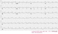
ECG of a patiënt with pulmonary embolism
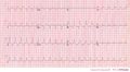
Another example of an ECG of a patiënt with pulmonary embolism. Note the tachycardia and right axis.
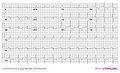
An example of right ventricular hypertrophy (and right atrial enlargement) in a patient with chronic pulmonary hypertension due to peripheral embolisation.
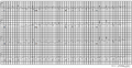
A 12 lead ECG of a patient with pulmonary embolism


