McGill Case 66: Difference between revisions
Jump to navigation
Jump to search
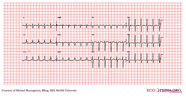
(Created page with "{{McGillcase| |previouspage= McGill Case 65 |previousname= McGill Case 65 |nextpage= McGill Case 67 |nextname= McGill Case 67 }} [[File:E0007661.jpg|thumb|600px|left|The firs...") |
No edit summary |
||
| Line 9: | Line 9: | ||
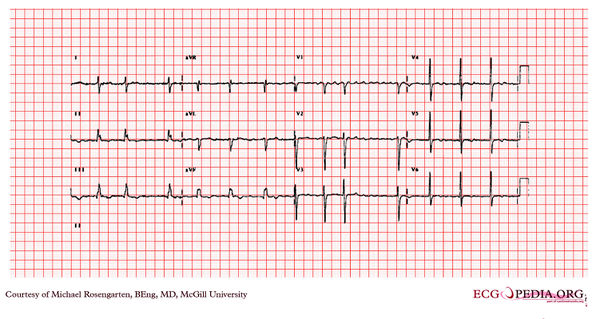
[[File:E0007662.jpg|thumb|600px|left|This tracing was taken in the intensive care unit after a temporary pacing wire (soft semi-floater) was placed via the right internal jugular vein. The lead paced the ventricle well, but the patient immediately complained of moderate chest pain, better with sitting up.]] | [[File:E0007662.jpg|thumb|600px|left|This tracing was taken in the intensive care unit after a temporary pacing wire (soft semi-floater) was placed via the right internal jugular vein. The lead paced the ventricle well, but the patient immediately complained of moderate chest pain, better with sitting up.]] | ||
Revision as of 01:03, 15 February 2012
| This case report is kindly provided by Michael Rosengarten from McGill and is part of the McGill Cases. These cases come from the McGill EKG World Encyclopedia.
|


