McGill Case 323: Difference between revisions
Jump to navigation
Jump to search
(Created page with "{{McGillcase| |previouspage= McGill Case 322 |previousname= McGill Case 322 |nextpage= McGill Case 325 |nextname= McGill Case 325 }} [[File:E323.jpg|thumb|600px|left|This is ...") |
No edit summary |
||
| Line 2: | Line 2: | ||
|previouspage= McGill Case 322 | |previouspage= McGill Case 322 | ||
|previousname= McGill Case 322 | |previousname= McGill Case 322 | ||
|nextpage= McGill Case | |nextpage= McGill Case 324 | ||
|nextname= McGill Case | |nextname= McGill Case 324 | ||
}} | }} | ||
Latest revision as of 23:06, 19 February 2012
|

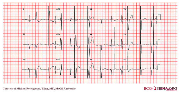
This is an electrocardiogram taken of from a patient after implantation of pacemaker. This electrocardiogram is interesting as it comes from a patient who previously had had a ventricular lead pacing his left ventricle. In this recording one clearly sees a QRS which now shows a left a bundle branch like configuration, suggesting that the New lead is in the right ventricle. One can also see that the patient has what is called trifasicular block, as we can see a first-degree block, a left axis deviation, and a right to bundle branch block. It is clear, that the pacemaker is not sensing the atrium or the ventricle. In this case, this is because a magnet was place over the pacemaker. This was required to demonstrate the QRS morphology from the paced beats as the patient's own heart rate was faster than the rate of the pacemaker.