McGill Case 217: Difference between revisions
Jump to navigation
Jump to search
(Created page with "{{McGillcase| |previouspage= McGill Case 216 |previousname= McGill Case 216 |nextpage= McGill Case 218 |nextname= McGill Case 218 }} [[File:E218.jpg|thumb|600px|left|The rhyt...") |
No edit summary |
||
| Line 6: | Line 6: | ||
}} | }} | ||
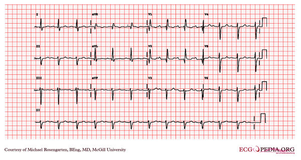
[[File: | [[File:E217.jpg|thumb|600px|left|The rhythm is sinus with a first degree heart block (pr > 120ms), there is a wide QRS (>120ms.) with a RBBB morphology and a left axis deviation consistent with a left fasicular block. The combination has been called a "trifasicular block" suggesting that the prolonged pr interval is due to slowing in the left posterior fascicle, but in fact this can be due to slowed conduction in the A/V node.]] | ||
Latest revision as of 05:21, 21 February 2012
|

The rhythm is sinus with a first degree heart block (pr > 120ms), there is a wide QRS (>120ms.) with a RBBB morphology and a left axis deviation consistent with a left fasicular block. The combination has been called a "trifasicular block" suggesting that the prolonged pr interval is due to slowing in the left posterior fascicle, but in fact this can be due to slowed conduction in the A/V node.