McGill Case 18: Difference between revisions
Jump to navigation
Jump to search
(Created page with "{{McGillcase| |previouspage= McGill Case 17 |previousname= McGill Case 17 |nextpage= McGill Case 19 |nextname= McGill Case 19 }} [[File:E0005605.jpg|thumb|600px|left|In a 41 ...") |
No edit summary |
||
| (One intermediate revision by the same user not shown) | |||
| Line 6: | Line 6: | ||
}} | }} | ||
[[File: | [[File:E000718.jpg|thumb|600px|left|This is an electrocardiogram from a woman in her forties who had several operations for congenital heart disease. At the time of the electrocardiogram the patient was taking flecainide and metoprolol. | ||
This patient was being treated for ventricular tachycardia. She was initially treated with amiodarone and then was switched to a combination of flecainide and metoprolol. She was doing well. The underlying congenital heart disease was Tetralolgy of Fallot. | |||
The electrocardiogram shows a supraventricular rhythm which is probably not sinus as indicated by the negative P waves in the inferior leads. The cardiogram also shows 1 PVC and a right Branch block with a left anterior hemi-block. | |||
.]] | |||
Latest revision as of 05:20, 10 February 2012
|

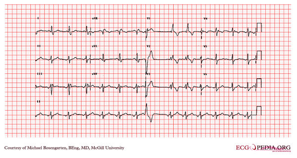
This is an electrocardiogram from a woman in her forties who had several operations for congenital heart disease. At the time of the electrocardiogram the patient was taking flecainide and metoprolol. This patient was being treated for ventricular tachycardia. She was initially treated with amiodarone and then was switched to a combination of flecainide and metoprolol. She was doing well. The underlying congenital heart disease was Tetralolgy of Fallot. The electrocardiogram shows a supraventricular rhythm which is probably not sinus as indicated by the negative P waves in the inferior leads. The cardiogram also shows 1 PVC and a right Branch block with a left anterior hemi-block. .