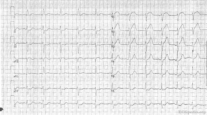
MI 2: Difference between revisions
Jump to navigation
Jump to search
m (New page: Culprit lesion: '''Left main''' # Sinus rhythm # Around 75 bpm # PQ normal, QRS interval 0.11 seconde, QT not prolonged # Left axis # normal p wave # QRS morphology: widened QRS, but not ...) |
mNo edit summary |
||
| (One intermediate revision by the same user not shown) | |||
| Line 1: | Line 1: | ||
{{Case| | |||
|previouspage= MI 1 | |||
|previousname= MI 1 | |||
|nextpage=MI 3 | |||
|nextname=MI 3 | |||
}} | |||
'''Where is this myocardial infarction located?''' | |||
[[Image:ami0002.jpg|700px|thumb|left|ECG MI 2]] | |||
{{clr}} | |||
[[Answer MI 2|Answer]] | |||
Latest revision as of 09:14, 11 November 2008
| This page is part of Cases and Examples |
Where is this myocardial infarction located?
