File:E365.jpg
Original file (3,004 × 1,599 pixels, file size: 4.44 MB, MIME type: image/jpeg)
Summary
| Description |
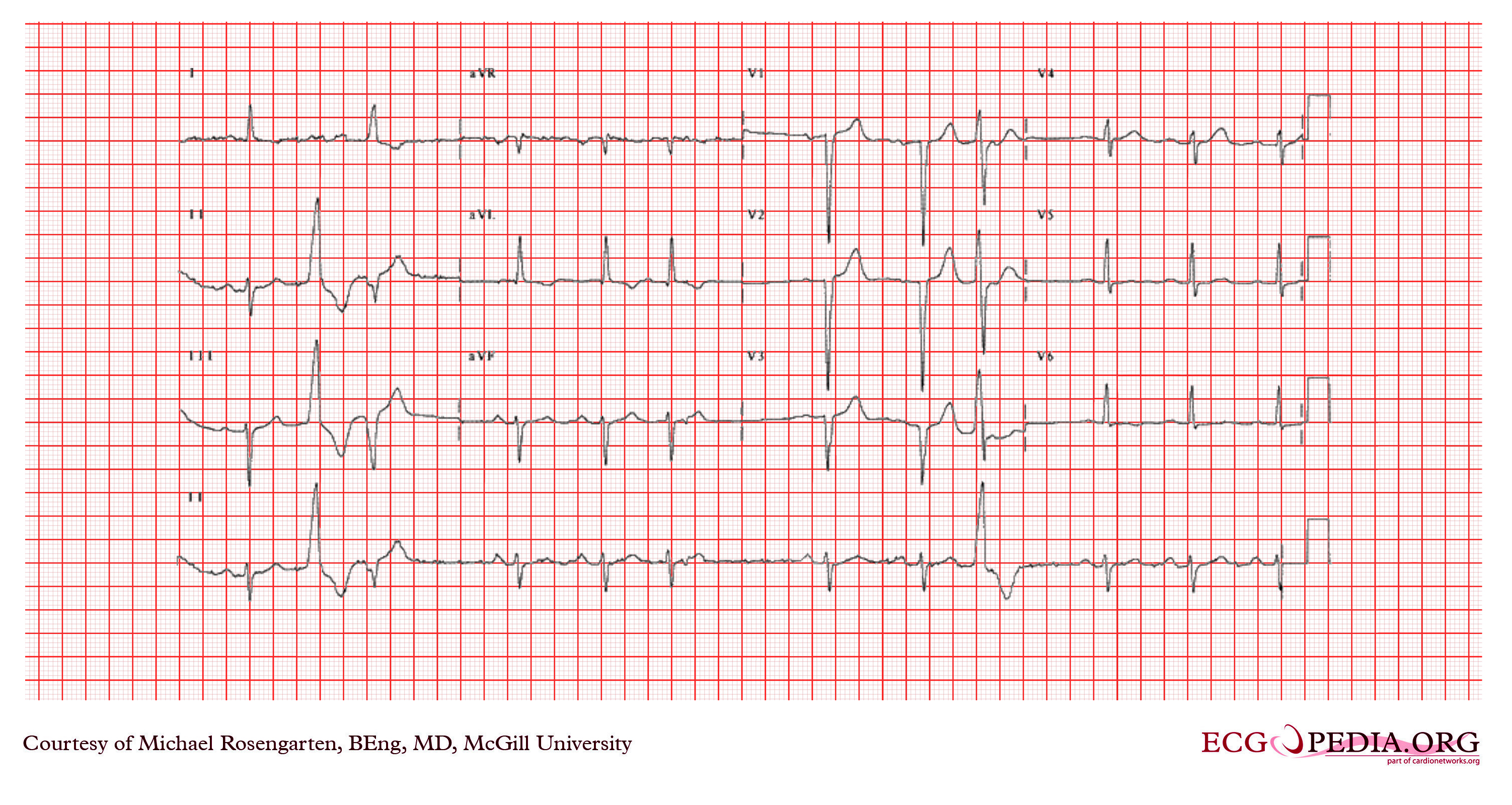
The rhythm is sinus rhythm at about 65/minute, with a PR interval of 200 ms. The QRS has a left axis deviation. There is a poor R wave progression across precordial leads. This suggests a left aterior hemi-block and a possible remote aterior wall myocardial infarction. There is also a multiform ventricular couplet (second and third beat) as well as a blocked PAC seen in the ST segment of the 6th beat. The 9th beat is a ventricular premature beat. |
|---|---|
| Category | |
| Source |
EKG World Encyclopedia http://cme.med.mcgill.ca/php/index.php , courtesy of Michael Rosengarten BEng, MD.McGill |
| Date |
2012 |
| Author |
Michael Rosengarten BEng, MD.McGill |
| Permission |
Creative Commons Attribution Noncommercial Share-Alike License |
File history
Click on a date/time to view the file as it appeared at that time.
| Date/Time | Thumbnail | Dimensions | User | Comment | |
|---|---|---|---|---|---|
| current | 11:50, 21 February 2012 | | 3,004 × 1,599 (4.44 MB) | DarrelC (talk | contribs) |
You cannot overwrite this file.
File usage
The following page uses this file: