File:E357.jpg
Jump to navigation
Jump to search
Size of this preview: 800 × 426 pixels. Other resolution: 3,004 × 1,599 pixels.
Original file (3,004 × 1,599 pixels, file size: 4.46 MB, MIME type: image/jpeg)
Summary
| Description |
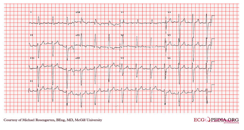
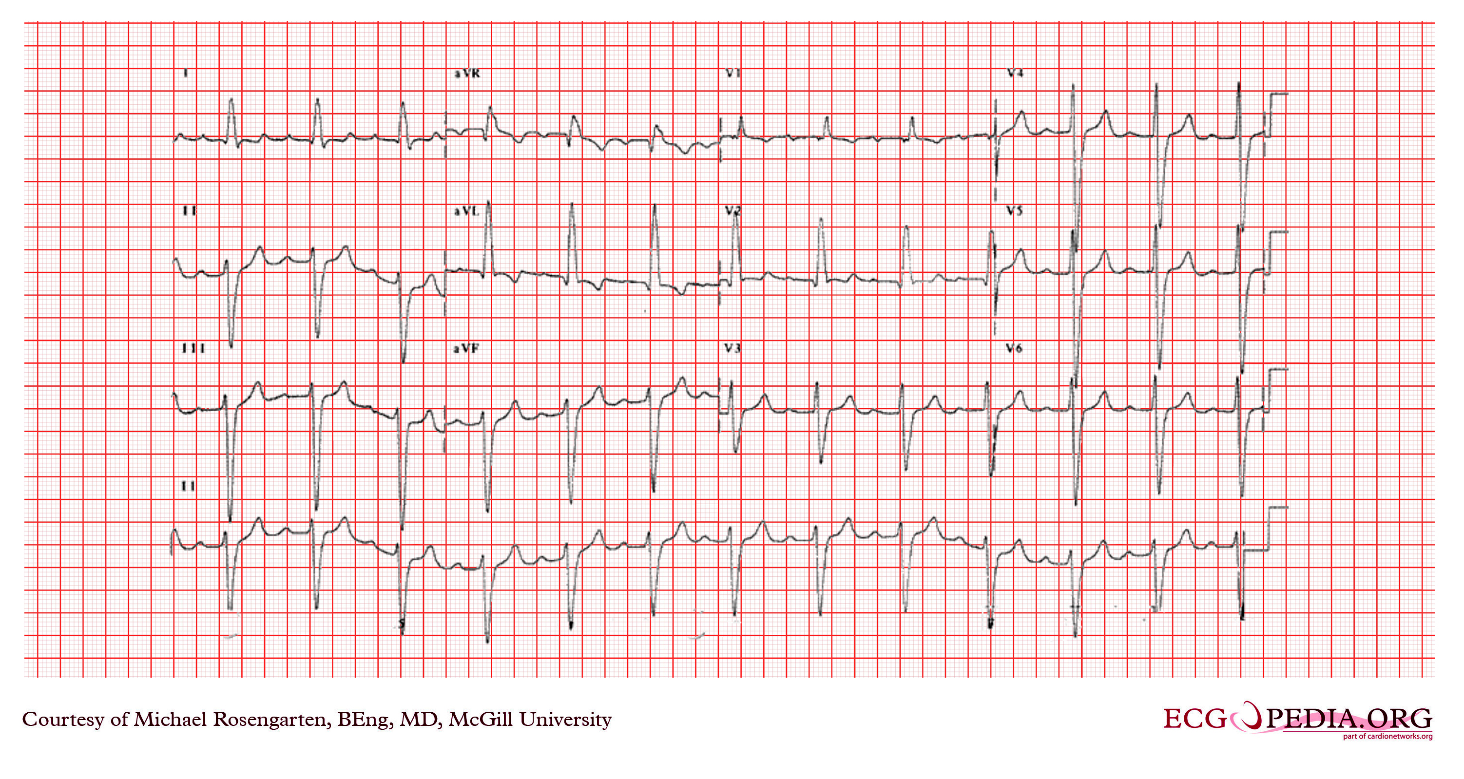
This is a regular rhythm with a ventricular rate of 70/min. The QRS has a left axis deviation and a dominant R wave in V1. The PR interval is 260 ms. This tracing suggests a first degree heart block with a left anterior hemi block and RBBB. This has been called Trifasicular block. |
|---|---|
| Category | |
| Source |
EKG World Encyclopedia http://cme.med.mcgill.ca/php/index.php , courtesy of Michael Rosengarten BEng, MD.McGill |
| Date |
2012 |
| Author |
Michael Rosengarten BEng, MD.McGill |
| Permission |
Creative Commons Attribution Noncommercial Share-Alike License |
File history
Click on a date/time to view the file as it appeared at that time.
| Date/Time | Thumbnail | Dimensions | User | Comment | |
|---|---|---|---|---|---|
| current | 11:36, 21 February 2012 | | 3,004 × 1,599 (4.46 MB) | DarrelC (talk | contribs) |
You cannot overwrite this file.
File usage
The following page uses this file: