File:E342.jpg
Original file (3,004 × 1,599 pixels, file size: 4.48 MB, MIME type: image/jpeg)
Summary
| Description |
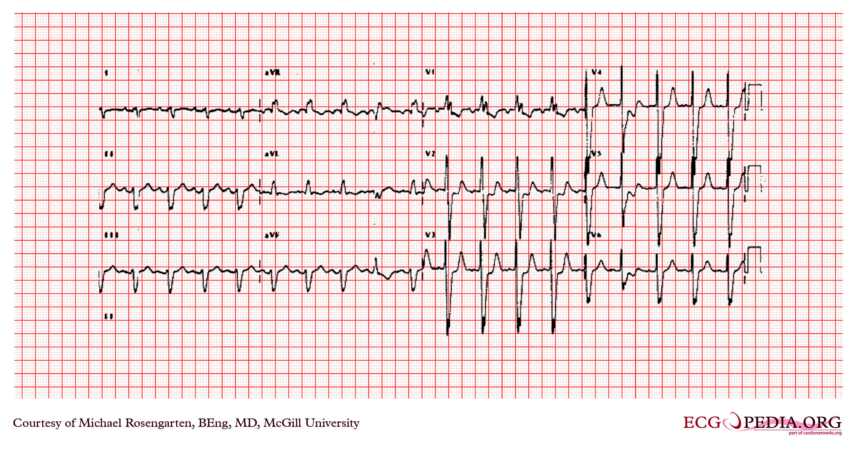
This is a recording from an older man in the surgical intensive care unit. He was recovering from a motor vehicle accident where he sustained a chest injury from his seat belt. The rhythm is sinus rhythm with a prolonged p wave duration in lead III( >140ms) and a pronounced terminal negativity in V1 suggtestive of left atrial abnormality. The QRS is wide with a duration of 137ms and a superior and right ward axis. There is an Rsr' in V1 ahd the S wave is greater than the R in V6. This is an unusual pattern for aberrance and is more in keeping with ventricular ectopy. In this case thought this appears to be a right bundle branch block with a possible left posterior hemi-block. Of note, in spite of this conduction disturbance the patient was able to sustain reentrant supraventricular tachycaridas requiring intravenous adenosine for termination. |
|---|---|
| Category | |
| Source |
EKG World Encyclopedia http://cme.med.mcgill.ca/php/index.php , courtesy of Michael Rosengarten BEng, MD.McGill |
| Date |
2012 |
| Author |
Michael Rosengarten BEng, MD.McGill |
| Permission |
Creative Commons Attribution Noncommercial Share-Alike License |
File history
Click on a date/time to view the file as it appeared at that time.
| Date/Time | Thumbnail | Dimensions | User | Comment | |
|---|---|---|---|---|---|
| current | 10:51, 21 February 2012 | | 3,004 × 1,599 (4.48 MB) | DarrelC (talk | contribs) |
You cannot overwrite this file.
File usage
The following page uses this file: