File:E330.jpg

Original file (3,004 × 1,599 pixels, file size: 4.47 MB, MIME type: image/jpeg)
Summary
| Description |
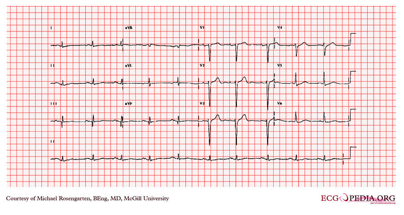
This is an electrocardiogram, a 64 year-old man with a history of coronary artery disease. The patient was taking amiodarone , metoprolol , and Vasotec at the time of this recording. This was a routine recording. The electrocardiogram shows sinus rhythm with a prolonged P wave > 120 milliseconds. The P wave is notched in the inferior leads. This is consistent with a left atrial abnormality. The PR interval is long at 200 milliseconds and is diagnostic of first-degree heart block. The Q waves in the Interior leads suggest an inferior wall infarction . |
|---|---|
| Category | |
| Source |
EKG World Encyclopedia http://cme.med.mcgill.ca/php/index.php , courtesy of Michael Rosengarten BEng, MD.McGill |
| Date |
2012 |
| Author |
Michael Rosengarten BEng, MD.McGill |
| Permission |
Creative Commons Attribution Noncommercial Share-Alike License |
File history
Click on a date/time to view the file as it appeared at that time.
| Date/Time | Thumbnail | Dimensions | User | Comment | |
|---|---|---|---|---|---|
| current | 10:28, 21 February 2012 | | 3,004 × 1,599 (4.47 MB) | DarrelC (talk | contribs) |
You cannot overwrite this file.
File usage
The following page uses this file: