File:E326.jpg

Original file (3,004 × 1,599 pixels, file size: 4.47 MB, MIME type: image/jpeg)
Summary
| Description |
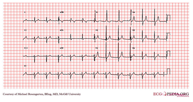
This is an electrocardiogram from a man in his 80's. The patient has severe lung disease, has mitral regurgitation secondary to bacterial endocarditis , and is taking digoxin, Lasix and potassium. The electrocardiogram shows sinus rhythm and a QRS with a left axis deviation, a QRS duration of 118 milliseconds and a tall are wave in the first precordial lead V1 with an R wave height of approximately 21 mm. The prolonged QRS duration and the S waves that are seen as lead one and lead the six suggest a right on the Branch block and the a left axis deviation suggests a left anterior hemi-block . Finally the tall R wave in V1 lead suggests right to ventricular Hypertrophic. |
|---|---|
| Category | |
| Source |
EKG World Encyclopedia http://cme.med.mcgill.ca/php/index.php , courtesy of Michael Rosengarten BEng, MD.McGill |
| Date |
2012 |
| Author |
Michael Rosengarten BEng, MD.McGill |
| Permission |
Creative Commons Attribution Noncommercial Share-Alike License |
File history
Click on a date/time to view the file as it appeared at that time.
| Date/Time | Thumbnail | Dimensions | User | Comment | |
|---|---|---|---|---|---|
| current | 10:10, 21 February 2012 | | 3,004 × 1,599 (4.47 MB) | DarrelC (talk | contribs) |
You cannot overwrite this file.
File usage
The following page uses this file: