File:E295.jpg
Original file (3,292 × 1,887 pixels, file size: 5.78 MB, MIME type: image/jpeg)
Summary
| Description |
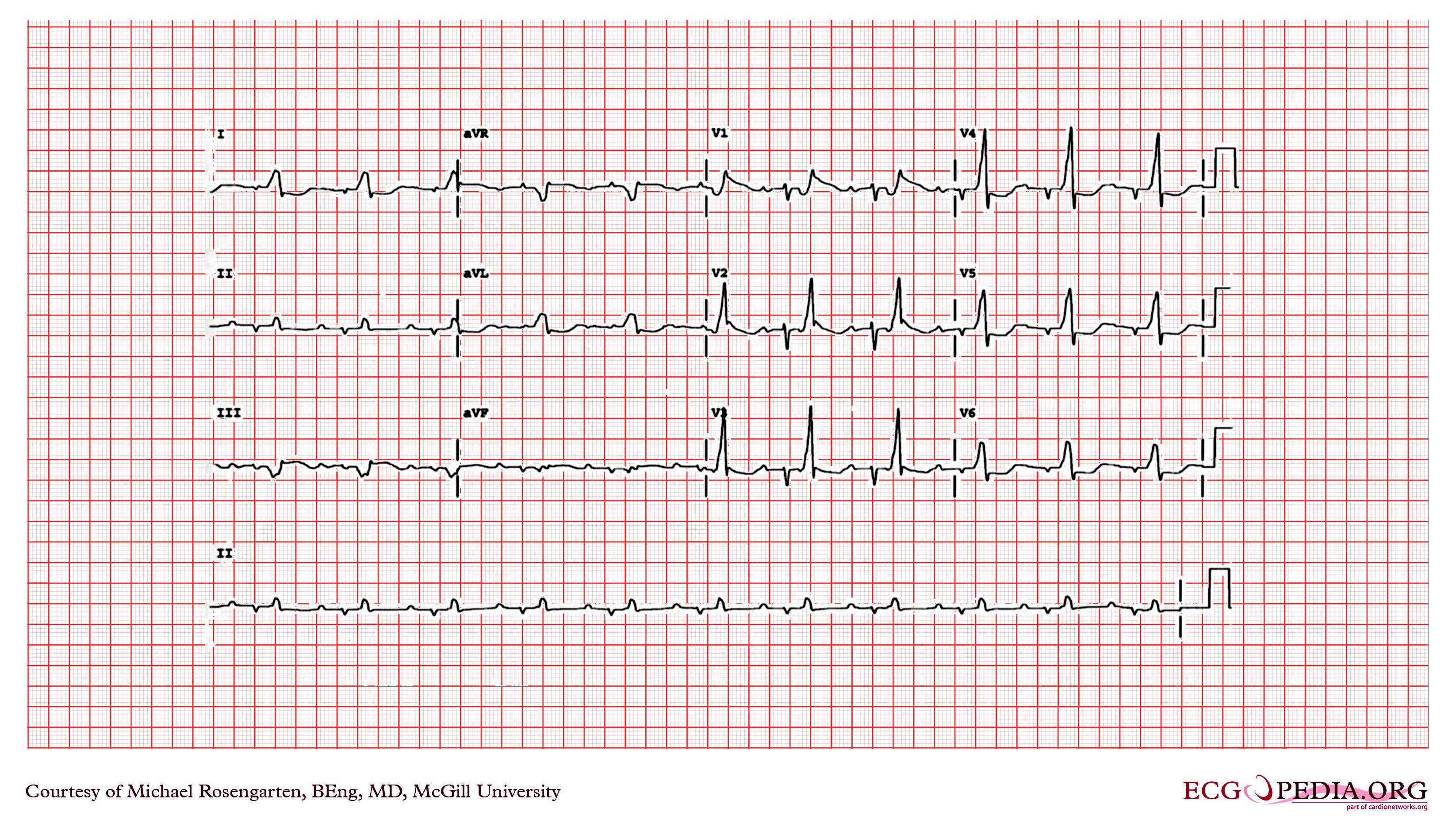
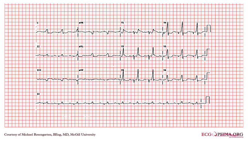
Intraventicular Conduction Defect comment: The ECG is in sinus rhythm and the QRS is markedly widened with a QRS duration of 260ms. The QRS seems split and gives the impression of ventricular bigemini but note that the second QRS deflection that looks like a PVC is in fact 200 ms after the onset of the first part of the QRS and hence too early for a PVC. Of interest this patient has recurrent ventricular tachycardia which may relate to his grossly widened QRS. The progression of the block can be seen over a three year period. comment from the web: When presented as a puzzler the correct interpretation of this ECG was not received, only suggestions of ventricular bigemini where given. |
|---|---|
| Category | |
| Source |
EKG World Encyclopedia http://cme.med.mcgill.ca/php/index.php , courtesy of Michael Rosengarten BEng, MD.McGill |
| Date |
2012 |
| Author |
Michael Rosengarten BEng, MD.McGill |
| Permission |
Creative Commons Attribution Noncommercial Share-Alike License |
File history
Click on a date/time to view the file as it appeared at that time.
| Date/Time | Thumbnail | Dimensions | User | Comment | |
|---|---|---|---|---|---|
| current | 07:47, 21 February 2012 | | 3,292 × 1,887 (5.78 MB) | DarrelC (talk | contribs) |
You cannot overwrite this file.
File usage
The following page uses this file: