File:E29.jpg

Original file (3,004 × 1,599 pixels, file size: 4.53 MB, MIME type: image/jpeg)
Summary
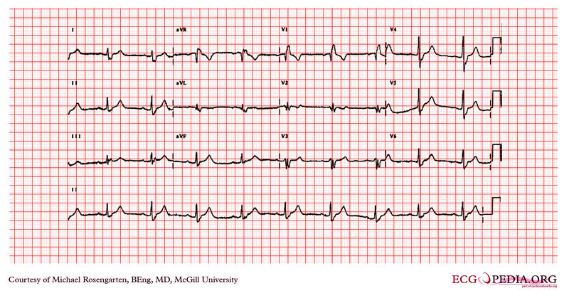
| Description |
This is an electrocardiogram from an elderly woman who had previously undergone surgery for recurrent ventricular tachycardia. She was being treated with Tambacor and metoprolol. The cardiogram shows sinus rhythm with a wide QRS of 159ms consistent with a RBBB and a rightward axis suggesting right posterior hemi-block. The PR interval is slightly prolonged at 2121ms. The poor R wave progression seen best in lead V2 suggests previous anterior wall MI. |
|---|---|
| Category | |
| Source |
EKG World Encyclopedia http://cme.med.mcgill.ca/php/index.php , courtesy of Michael Rosengarten BEng, MD.McGill |
| Date |
2012 |
| Author |
Michael Rosengarten BEng, MD.McGill |
| Permission |
Creative Commons Attribution Noncommercial Share-Alike License |
File history
Click on a date/time to view the file as it appeared at that time.
| Date/Time | Thumbnail | Dimensions | User | Comment | |
|---|---|---|---|---|---|
| current | 10:27, 21 February 2012 | | 3,004 × 1,599 (4.53 MB) | DarrelC (talk | contribs) |
You cannot overwrite this file.
File usage
The following page uses this file: