File:E276.jpg
Jump to navigation
Jump to search
Size of this preview: 799 × 196 pixels. Other resolution: 3,004 × 737 pixels.
Original file (3,004 × 737 pixels, file size: 806 KB, MIME type: image/jpeg)
Summary
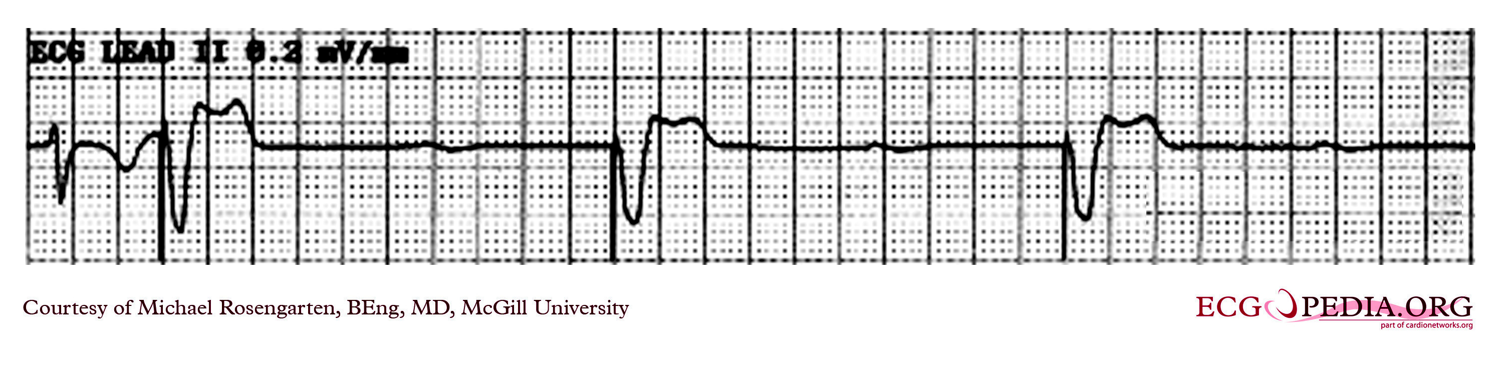
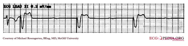
| Description |
The recording shows one native ventricular complex followed by three paced venticular beats. Sinus P waves can be seen floating through and unrelated to the ventricular complexes. There is no evidence of conduction from the atrium. |
|---|---|
| Category | |
| Source |
EKG World Encyclopedia http://cme.med.mcgill.ca/php/index.php , courtesy of Michael Rosengarten BEng, MD.McGill |
| Date |
2012 |
| Author |
Michael Rosengarten BEng, MD.McGill |
| Permission |
Creative Commons Attribution Noncommercial Share-Alike License |
File history
Click on a date/time to view the file as it appeared at that time.
| Date/Time | Thumbnail | Dimensions | User | Comment | |
|---|---|---|---|---|---|
| current | 07:09, 21 February 2012 | 3,004 × 737 (806 KB) | DarrelC (talk | contribs) |
You cannot overwrite this file.
File usage
The following page uses this file: