File:E268.jpg
Original file (3,004 × 1,599 pixels, file size: 4.43 MB, MIME type: image/jpeg)
Summary
| Description |
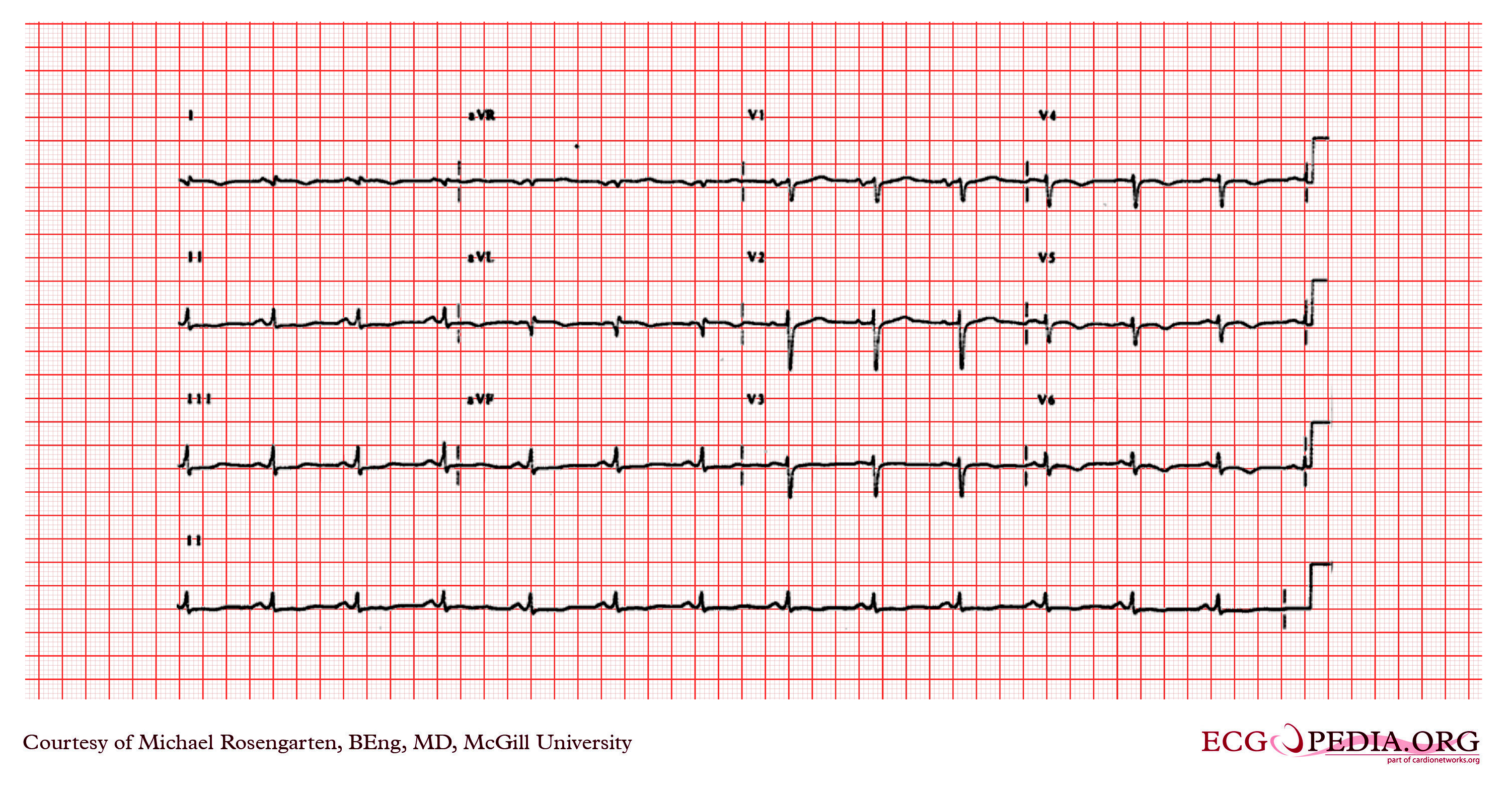
The cardiogram shows sinus rhythm and a QRS with a rightward axis, as well as wide Q waves in leads I and AVL as well as a poor r wave progression across the anterior chest leads. There is also slight ST elevation in leads I, aVL, and T wave inversion in the lateral leads. The EKG is consistent with a lateral wall myocardial infarction. The patient had had a myocardial infarction a few months before. This event was associated with a cardiac arrest due to ventricular fibrillation which was successfully treated by the 911 ambulance service. |
|---|---|
| Category | |
| Source |
EKG World Encyclopedia http://cme.med.mcgill.ca/php/index.php , courtesy of Michael Rosengarten BEng, MD.McGill |
| Date |
2012 |
| Author |
Michael Rosengarten BEng, MD.McGill |
| Permission |
Creative Commons Attribution Noncommercial Share-Alike License |
File history
Click on a date/time to view the file as it appeared at that time.
| Date/Time | Thumbnail | Dimensions | User | Comment | |
|---|---|---|---|---|---|
| current | 06:53, 21 February 2012 | | 3,004 × 1,599 (4.43 MB) | DarrelC (talk | contribs) |
You cannot overwrite this file.
File usage
The following page uses this file: