File:E263.jpg
Original file (3,004 × 1,599 pixels, file size: 4.43 MB, MIME type: image/jpeg)
Summary
| Description |
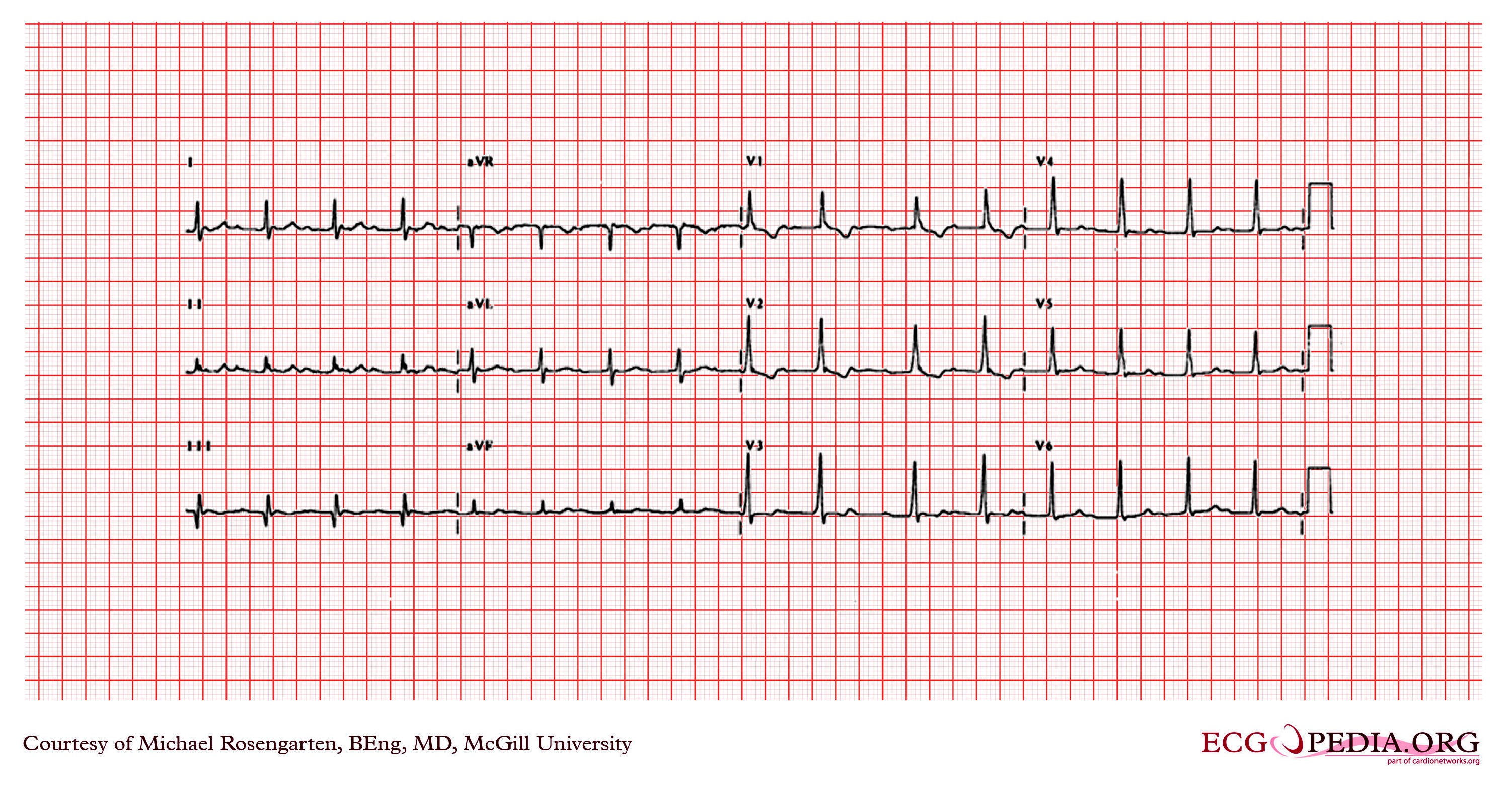
This is a recording from a patient being treated for a wide complex tachycardia . The recording could be sinus with a first degree block (plus a partial right bundle branch block or possibly RVH). It is to be noted though that a sinus rate of 100/min seems some what fast for a patient on nadolol and that the rhythm is irregular. On the other hand, the more likely rhyhtm is SVT (or slow atrial flutter at a rate of 200/min) with variable block though mostly 2:1 block. This is an atrial rate similar to that of the wide complex tachycardia, and if one looks in lead I and lead V1 there does appear to be atrial activity at twice the ventricular rate. |
|---|---|
| Category | |
| Source |
EKG World Encyclopedia http://cme.med.mcgill.ca/php/index.php , courtesy of Michael Rosengarten BEng, MD.McGill |
| Date |
2012 |
| Author |
Michael Rosengarten BEng, MD.McGill |
| Permission |
Creative Commons Attribution Noncommercial Share-Alike License |
File history
Click on a date/time to view the file as it appeared at that time.
| Date/Time | Thumbnail | Dimensions | User | Comment | |
|---|---|---|---|---|---|
| current | 06:42, 21 February 2012 | | 3,004 × 1,599 (4.43 MB) | DarrelC (talk | contribs) |
You cannot overwrite this file.
File usage
The following page uses this file: