File:E201.jpg
Size of this preview: 800 × 426 pixels. Other resolution: 3,004 × 1,599 pixels.
Original file (3,004 × 1,599 pixels, file size: 4.43 MB, MIME type: image/jpeg)
Summary
| Description |
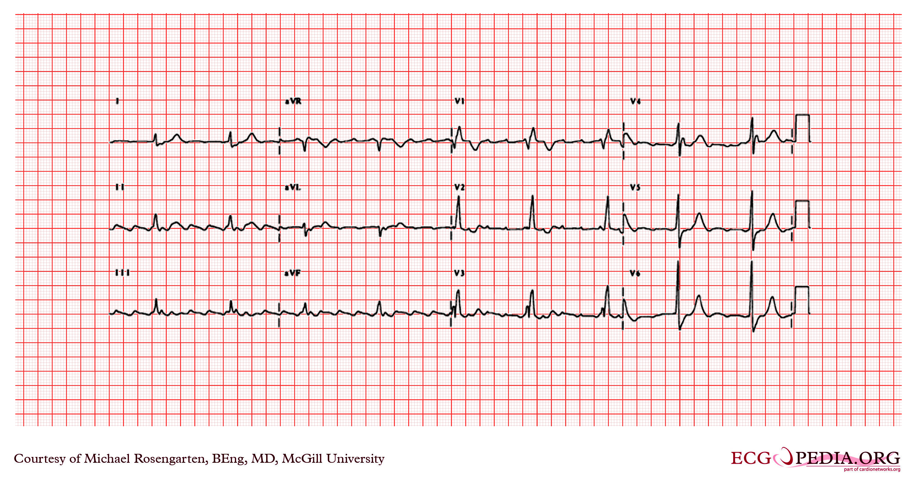
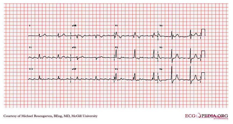
The rhythm is atrial flutter with flutter waves seen best in the inferior leads and in leads V1 to V3. The atrial rate is about 250/min. The QRS is wide (>120ms) an there is a tall R' wave in V1 and a shallow S in V6. The axis of the QRS seems normal. The EKG shows a right bundle branch block. |
|---|---|
| Category | |
| Source |
EKG World Encyclopedia http://cme.med.mcgill.ca/php/index.php , courtesy of Michael Rosengarten BEng, MD.McGill |
| Date |
2012 |
| Author |
Michael Rosengarten BEng, MD.McGill |
| Permission |
Creative Commons Attribution Noncommercial Share-Alike License |
File history
Click on a date/time to view the file as it appeared at that time.
| Date/Time | Thumbnail | Dimensions | User | Comment | |
|---|---|---|---|---|---|
| current | 04:00, 21 February 2012 | | 3,004 × 1,599 (4.43 MB) | DarrelC (talk | contribs) |
You cannot overwrite this file.
File usage
The following page uses this file: