File:E196.jpg
Original file (3,004 × 1,599 pixels, file size: 4.43 MB, MIME type: image/jpeg)
Summary
| Description |
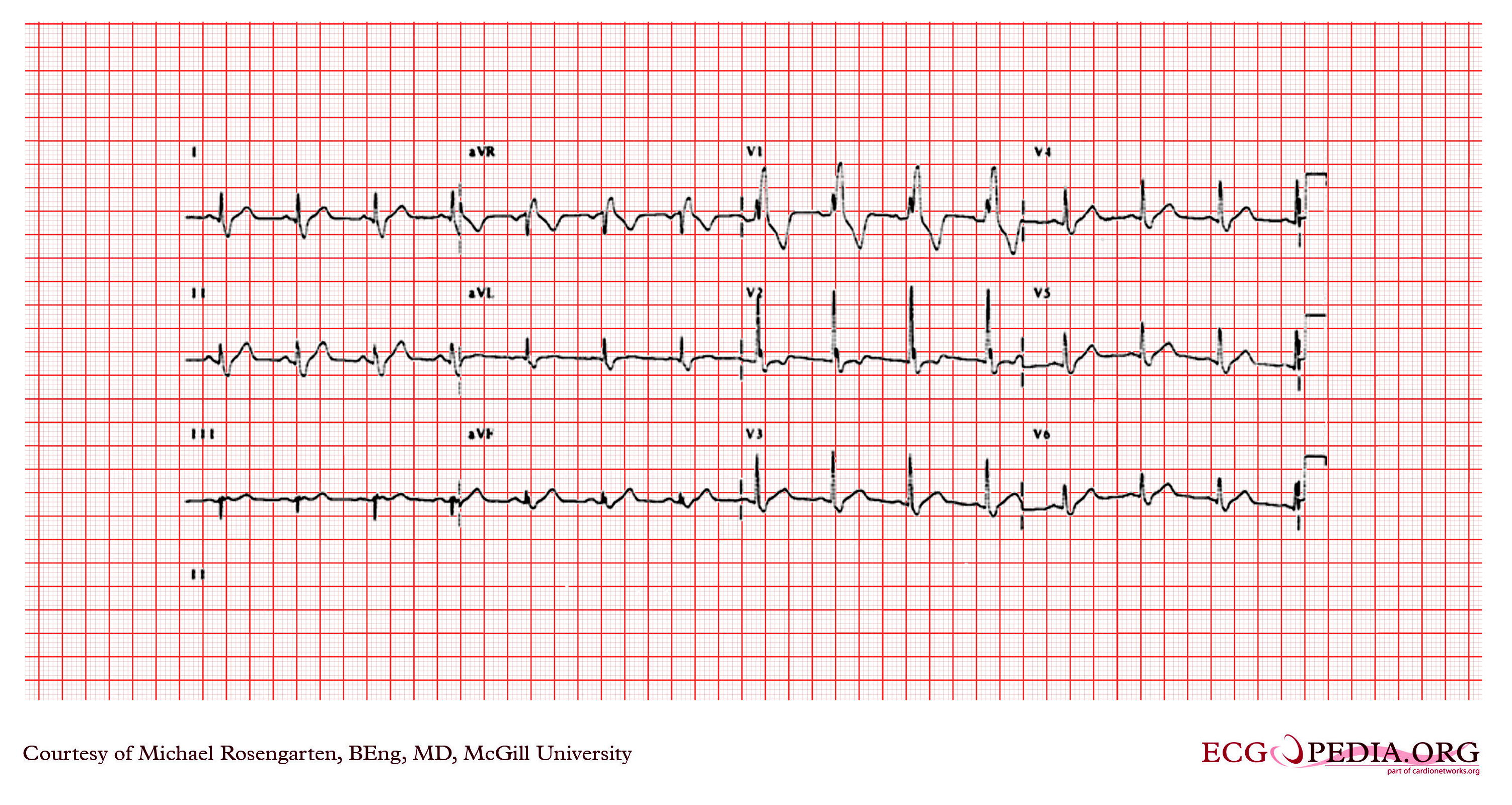
This cardiogram shows sinus rhythm with a normal pr interval and a prolonged QRS interval (>120ms). There is a conduction abnormality best described as a right bundle branch block due to the rsR' wave in V1. Note the S wave in V6 which is due to the RBBB is smaller than the R wave in V6. The axis of the QRS is difficult to determine, but one usually looks at the first 60 ms. (1 1/2 small squares) to determine the axis with a RBBB. If the axis of the first 60 ms. of the QRS is more than 90 degrees and there is an rS in lead I and a Q in lead III then on would consider a left posterior fasicular block. This is not the case here. |
|---|---|
| Category | |
| Source |
EKG World Encyclopedia http://cme.med.mcgill.ca/php/index.php , courtesy of Michael Rosengarten BEng, MD.McGill |
| Date |
2012 |
| Author |
Michael Rosengarten BEng, MD.McGill |
| Permission |
Creative Commons Attribution Noncommercial Share-Alike License |
File history
Click on a date/time to view the file as it appeared at that time.
| Date/Time | Thumbnail | Dimensions | User | Comment | |
|---|---|---|---|---|---|
| current | 00:25, 21 February 2012 | | 3,004 × 1,599 (4.43 MB) | DarrelC (talk | contribs) |
You cannot overwrite this file.
File usage
The following page uses this file: