File:E000789.jpg
Original file (3,004 × 1,599 pixels, file size: 2.24 MB, MIME type: image/jpeg)
Summary
| Description |
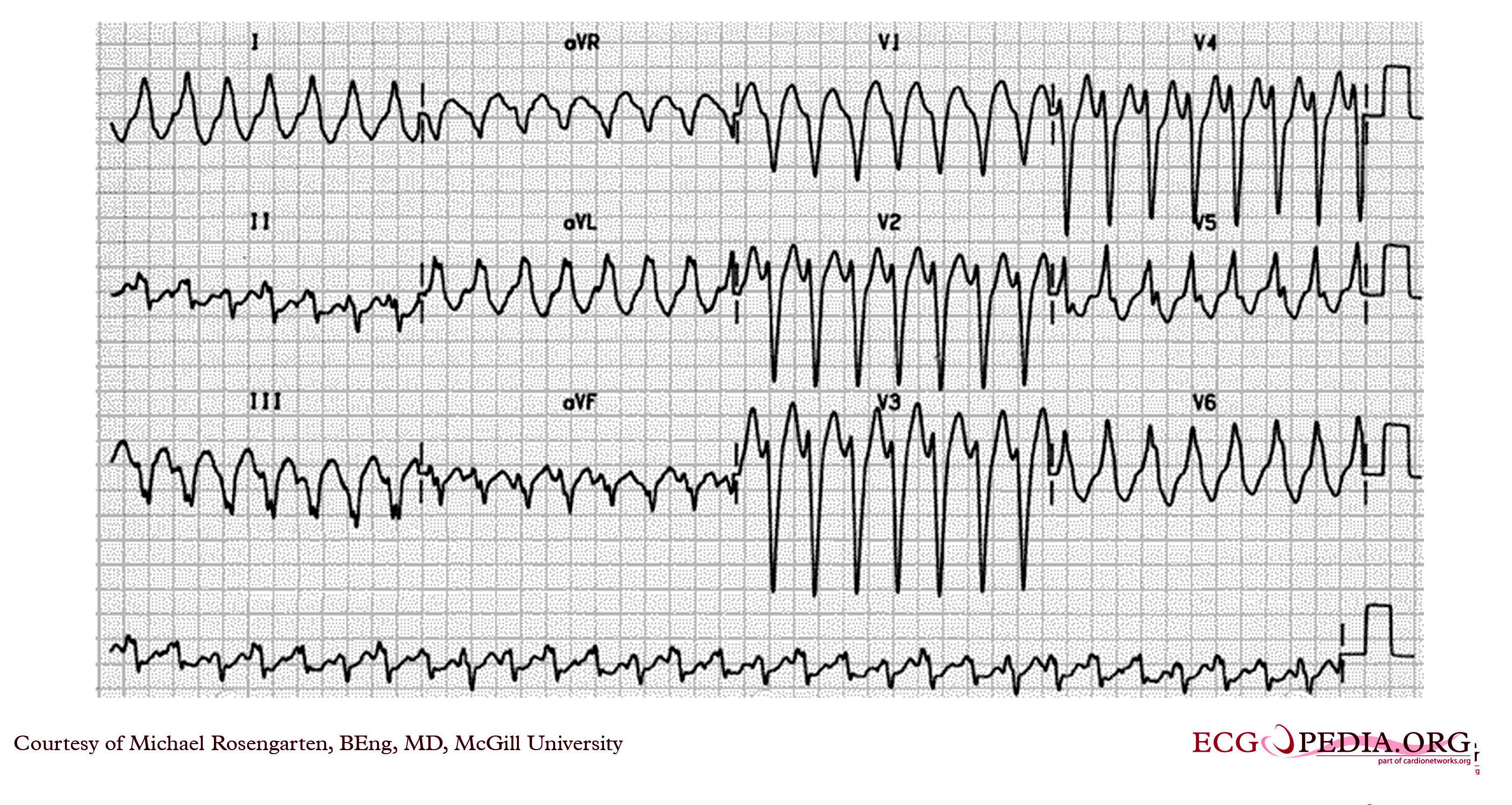
This is an electrocardiogram from an elderly woman with palpitations. The cardiogram shows a wide complex tachycardia with a left bundle branch morphology at a rate of about 160/min. The R wave in V2 is broad and the time from the beginning of the QRS in V2 to the peak of the S wave is longer than 80 ms. No P wave activity is clearly seen. The cardiogram suggests ventricular tachycardia. The patient has done well since this cardiogram on flecainide and metoprolol. |
|---|---|
| Category | |
| Source |
EKG World Encyclopedia http://cme.med.mcgill.ca/php/index.php , courtesy of Michael Rosengarten BEng, MD.McGill |
| Date |
2012 |
| Author |
Michael Rosengarten BEng, MD.McGill |
| Permission |
Creative Commons Attribution Noncommercial Share-Alike License |
File history
Click on a date/time to view the file as it appeared at that time.
| Date/Time | Thumbnail | Dimensions | User | Comment | |
|---|---|---|---|---|---|
| current | 04:44, 15 February 2012 | | 3,004 × 1,599 (2.24 MB) | DarrelC (talk | contribs) |
You cannot overwrite this file.
File usage
The following page uses this file: