File:E000734.jpg
Original file (3,004 × 1,599 pixels, file size: 4.44 MB, MIME type: image/jpeg)
Summary
| Description |
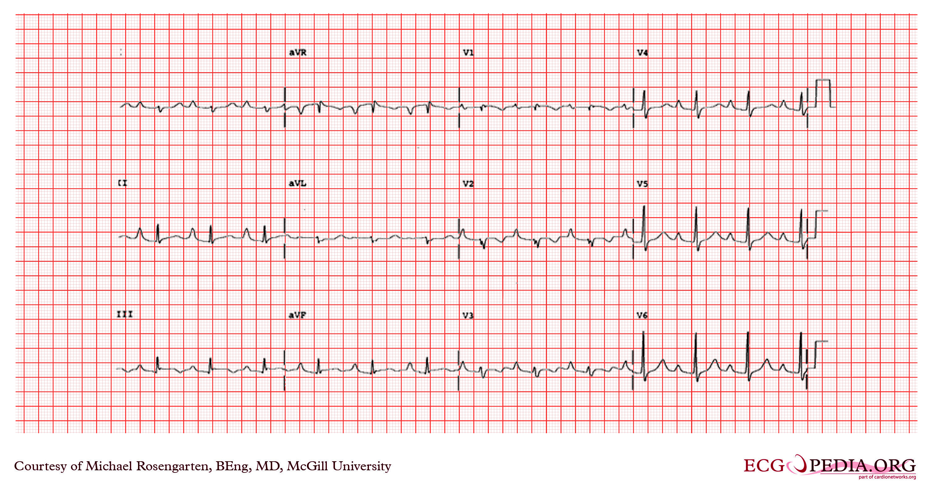
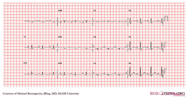
This is an electrocardiogram from a young woman with Ebstein's anomaly. In this condition the right atrium is enlarged with downward a displacement of the tricuspid valve into the right ventricle. This partially explains the tall peaked p waves in the inferior leads. Note the P wave in lead II is greater than 2 mm in height. There is also a first-degree heart block which is often seen with these patients. Curiously higher grade blocks are rare. The QRS complex shows a right axis deviation and a terminal S wave in the V 6 derivation. Atypical right bundle branch block can be seen with this condition. |
|---|---|
| Category | |
| Source |
EKG World Encyclopedia http://cme.med.mcgill.ca/php/index.php , courtesy of Michael Rosengarten BEng, MD.McGill |
| Date |
2012 |
| Author |
Michael Rosengarten BEng, MD.McGill |
| Permission |
Creative Commons Attribution Noncommercial Share-Alike License |
File history
Click on a date/time to view the file as it appeared at that time.
| Date/Time | Thumbnail | Dimensions | User | Comment | |
|---|---|---|---|---|---|
| current | 02:53, 11 February 2012 | | 3,004 × 1,599 (4.44 MB) | DarrelC (talk | contribs) |
You cannot overwrite this file.
File usage
The following page uses this file: