File:E0003174.jpg
Original file (3,004 × 634 pixels, file size: 674 KB, MIME type: image/jpeg)
Summary
| Description |
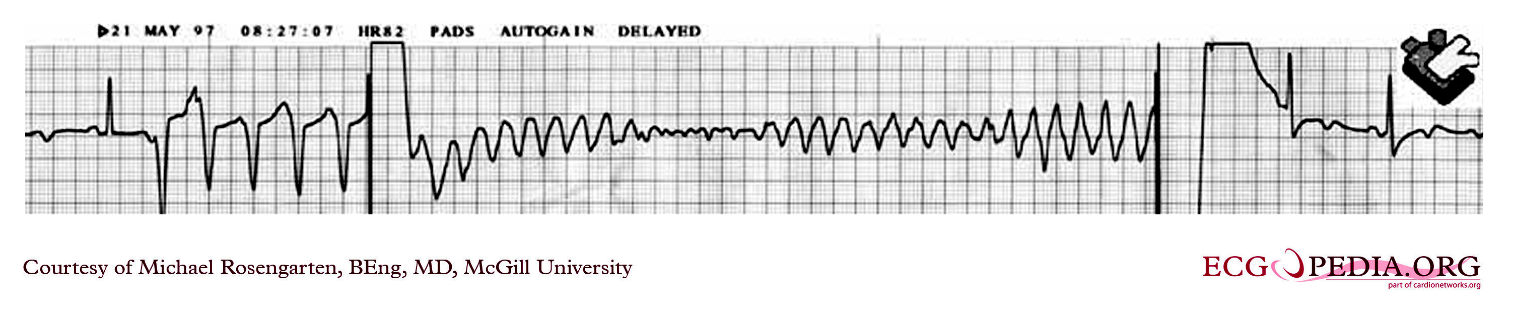
This is an EKG monitor strip recorded during the testing of a defibrillator in a middle aged woman with ventricular tachycardia and a structurally normal heart. The tracing was made during the implantation of the defibrillator and shows induction of VF with paced beats and a low energy T wave shock, termination of the VF by the device and then the return to sinus rhythm. Note the QT interval in this patient of 500 ms which is long for the heart rate of 60/min. |
|---|---|
| Category | |
| Source |
EKG World Encyclopedia http://cme.med.mcgill.ca/php/index.php , courtesy of Michael Rosengarten BEng, MD.McGill |
| Date |
2012 |
| Author |
Michael Rosengarten BEng, MD.McGill |
| Permission |
Creative Commons Attribution Noncommercial Share-Alike License |
File history
Click on a date/time to view the file as it appeared at that time.
| Date/Time | Thumbnail | Dimensions | User | Comment | |
|---|---|---|---|---|---|
| current | 01:21, 19 February 2012 | 3,004 × 634 (674 KB) | DarrelC (talk | contribs) |
You cannot overwrite this file.
File usage
The following page uses this file: