File:E0003123.jpg
Original file (3,004 × 1,599 pixels, file size: 4.47 MB, MIME type: image/jpeg)
Summary
| Description |
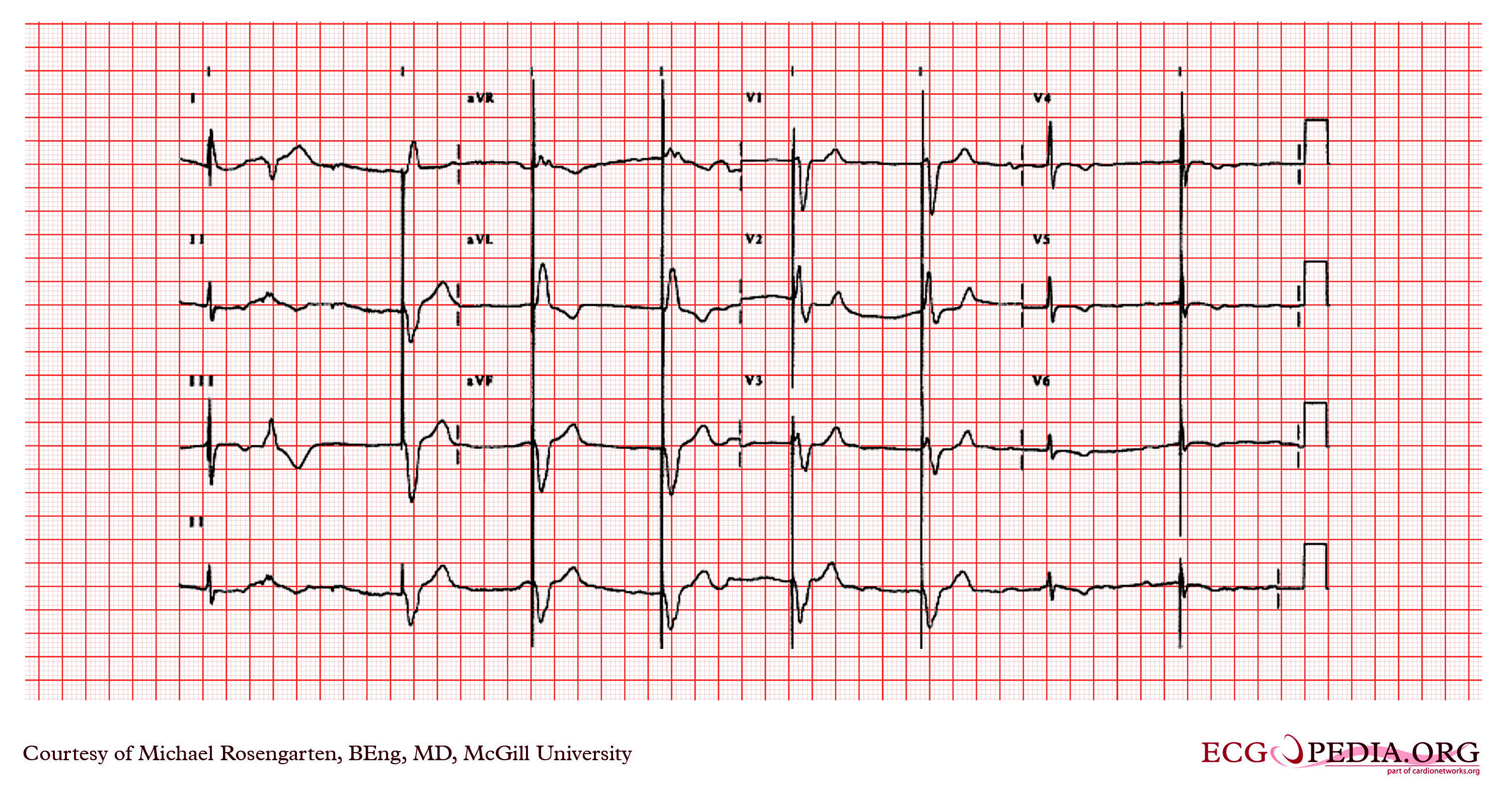
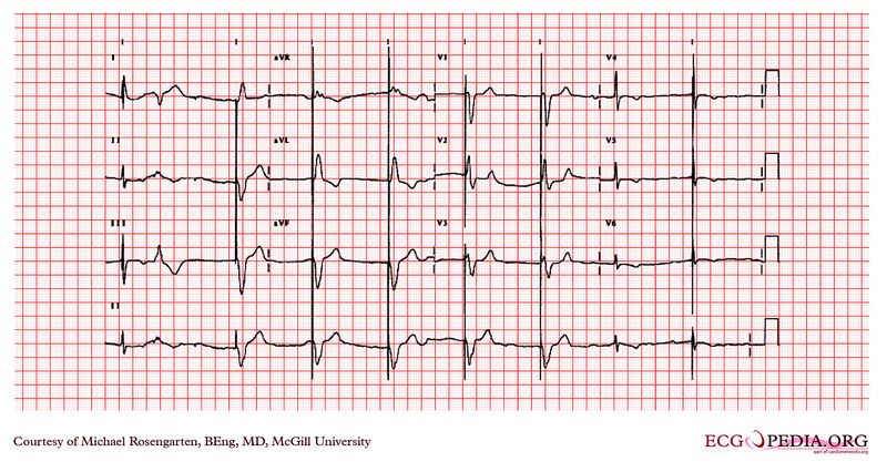
This is an electrocardiogram from a 87 year old man with a history of atrial fibrillation. His medications were coumadin and Monopril. The cardiogram shows sinus rhythm with rate of about 50/min, and a marked first degree heart block with a pr interval of about 350ms. The first complex on the left is a fusion between the patient's native QRS and the pacemaker spike (this is nomal operation) this is followed by a PVC. Note the small blip following the PVC is artifact and is not a failure to capture of the pacemaker. The pacemaker is working well as a VVI pacer set at 50/min. The large spikes suggest a unipolar lead. |
|---|---|
| Category | |
| Source |
EKG World Encyclopedia http://cme.med.mcgill.ca/php/index.php , courtesy of Michael Rosengarten BEng, MD.McGill |
| Date |
2012 |
| Author |
Michael Rosengarten BEng, MD.McGill |
| Permission |
Creative Commons Attribution Noncommercial Share-Alike License |
File history
Click on a date/time to view the file as it appeared at that time.
| Date/Time | Thumbnail | Dimensions | User | Comment | |
|---|---|---|---|---|---|
| current | 10:54, 17 February 2012 | | 3,004 × 1,599 (4.47 MB) | DarrelC (talk | contribs) |
You cannot overwrite this file.
File usage
The following page uses this file: