File:E.jpg: Difference between revisions
Jump to navigation
Jump to search
No edit summary |
No edit summary |
||
| Line 1: | Line 1: | ||
== Summary == | |||
{{Information | |||
|Description = These two strips from the same patient show a 2:1 block on the top tracing and a Mobitz II A/V block on the lower one. Note that with 2:1 block you can not tell if this is a Mobitz I or II. Mobitz II is seen below as the PR does not change before and after the non-conducted P wave. | |||
|Category = [[Case_reports_from_Michael_Rosengarten|Case reports by Michael Rosengarten]] | |||
|Source = EKG World Encyclopedia http://cme.med.mcgill.ca/php/index.php , courtesy of Michael Rosengarten BEng, MD.McGill | |||
|Date = 2012 | |||
|Author = Michael Rosengarten BEng, MD.McGill | |||
|Permission = {{by-nc-sa-3.0}} | |||
|other_versions = None | |||
}} | |||
Latest revision as of 06:58, 21 February 2012
Summary
| Description |
These two strips from the same patient show a 2:1 block on the top tracing and a Mobitz II A/V block on the lower one. Note that with 2:1 block you can not tell if this is a Mobitz I or II. Mobitz II is seen below as the PR does not change before and after the non-conducted P wave. |
|---|---|
| Category | |
| Source |
EKG World Encyclopedia http://cme.med.mcgill.ca/php/index.php , courtesy of Michael Rosengarten BEng, MD.McGill |
| Date |
2012 |
| Author |
Michael Rosengarten BEng, MD.McGill |
| Permission |
Creative Commons Attribution Noncommercial Share-Alike License |
File history
Click on a date/time to view the file as it appeared at that time.
| Date/Time | Thumbnail | Dimensions | User | Comment | |
|---|---|---|---|---|---|
| current | 06:57, 21 February 2012 | 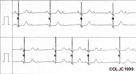 | 470 × 257 (17 KB) | DarrelC (talk | contribs) |
You cannot overwrite this file.
File usage
The following page uses this file: