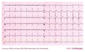
Example 24
Jump to navigation
Jump to search
- Following the 7+2 steps:
- Rhythm
- The ECG shows a regular rhythm. Every P wave is followed by a QRS complex. P waves are positive in I and AVF. Normal sinus rhythm
- Heart rate
- about 80bpm
- Conduction (PQ,QRS,QT)
- In lead II: PQ: 210ms QRS: 90ms QT: 380ms QTc: 450ms
- Heartaxis
- QRS positive in I and AVF: normal heart axis
- P wave morphology
- Broad based P waves
- QRS morphology
- Normal QRS complexes
- ST morphology
- Typical ST elevation in V1, V2 and V3, with a coved type morphology in V1.
- Compare with the old ECG (not available, so skip this step)
- Conclusion?
- Rhythm
Typical Brugada syndrome ST segments in right precordial ECG leads (on spot diagnosis) aka 'type-1 Brugada ECG' with 1st degree AV block and broad P-waves.
Atrial/AV/ventricular conduction delay is commonly seen in Brugada syndrome, in this patient there is atrial and AV conduction delay. Brugada syndrome is associated with malignant arrhythmias (VT/VF) and sudden death although many patients may not experience any symptoms throughout their life.
