Eponymous ECG's: Difference between revisions
m (→Osborn J wave) |
m (→Osborn J wave) |
||
| Line 18: | Line 18: | ||
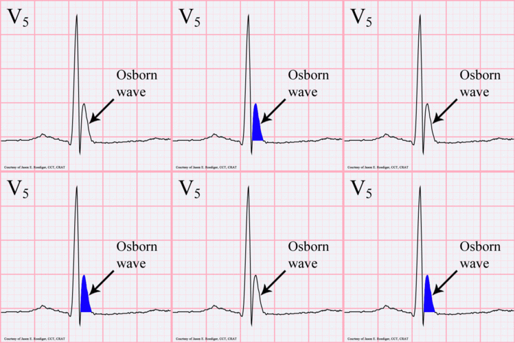
[[File:Osborn-wave.gif|250px|right|thumb|Osborn wave. 81-year-old black male with BP 80/62 and temperature 89.5 degrees F (32 C).]] | [[File:Osborn-wave.gif|250px|right|thumb|Osborn wave. 81-year-old black male with BP 80/62 and temperature 89.5 degrees F (32 C).]] | ||
The prominent J deflection attributed to hypothermia was first reported in 1938 by Tomaszewski. These waves were then definitively described in 1953 by John J. Osborn (born 1917) and were named in his honor.<cite>osborn</cite> | The prominent J deflection attributed to hypothermia was first reported in 1938 by Tomaszewski. These waves were then definitively described in 1953 by John J. Osborn (born 1917) and were named in his honor.<cite>osborn</cite> | ||
{{clr}} | |||
==References== | ==References== | ||
Latest revision as of 13:03, 15 January 2016
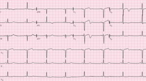
Wellens ECG
Wellens' ECG (sometimes referred to as Wellens syndrome or sign) is an ECG manifestation of proximal left anterior descending stenosis in patients with acute coronary syndrome. It is characterized by symmetrical, often deep (>2 mm), T wave inversions in the anterior precordial leads. A less common variant is biphasic T wave inversions in the same leads. [1]
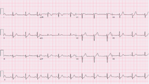
De Winter ECG
The De Winter ECG is sometimes seen in myocardial infarction with proximal LAD occlusion. It is rather rare (2% of cases). There is no anterior ST segment elevation. Instead the ST segment shows a 1- to 3-mm upsloping ST-segment depression at the J point in leads V1 to V6 that continues into tall, positive symmetrical T waves. The QRS complexes are usually not widened or only slightly widened, and sometimes there is a loss of precordial R-wave progression. In most patients there is a 1- to 2-mm ST-elevation in lead aVR. [2] Prof. Robert de Winter is an intervention cardiologist in the Academic Medical Center in Amsterdam, the Netherlands.
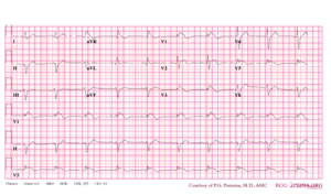
Brugada syndrome
The Brugada syndrome is an hereditary disease that is associated with high risk of sudden cardiac death. It is characterized by typical ECG abnormalities: ST segment elevation in the precordial leads (V1 - V3).[3]
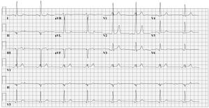
Wolff–Parkinson–White syndrome
In 1930 Louis Wolff, Sir John Parkinson and Paul Dudley White described 11 patients who suffered from bouts of tachcyardias. Their ECGs showed two abnormalities: a short PQ time and a delta-wave. Ever since one speaks of the Wolff-Parkinson-White syndrome in patients with complaints of syncope and / or tachycardia and a pre-exitation pattern on the ECG (WPW syndrome = WPW pattern + symptoms). Not all patients with a WPW pattern on the ECG are symptomatic. The prevalence of the WPW or pre-exitation pattern is relatively common in the general population (about 0.15-0.25%).[4] Template:Cld
Osborn J wave
The prominent J deflection attributed to hypothermia was first reported in 1938 by Tomaszewski. These waves were then definitively described in 1953 by John J. Osborn (born 1917) and were named in his honor.[5]
References
<biblio>
- dewinter pmid=18987380
- wellens pmid=6121481
- brugada pmid=1309182
- wpw pmid=17040283
- osborne pmid=15902836
</blblio>
