Cases and Examples: Difference between revisions
Jump to navigation
Jump to search
mNo edit summary |
No edit summary |
||
| Line 25: | Line 25: | ||
</gallery> | </gallery> | ||
=== | ===Rarities=== | ||
<gallery | <gallery> | ||
Image: | Image:ECG_Aflutt_1to1.jpg|Atrial flutter with 1:1 conduction | ||
Image:JEt.jpg|Junctional Ectopic Tachycardia (JEt) | |||
Image:pacemaker_retrograde_wenkebach.jpg|Pacemakerrhythm with retrograde Wenkebach. | |||
Image:T_wave_alternans.png|Extreme prolonged QT interval with T wave alternans | |||
</gallery> | </gallery> | ||
Revision as of 15:05, 23 July 2007
Below you can find some common examples. ECGs can be magnified by clicking on the image....
Simple
Click on the name below the ECG for the case descriptions. Click on the ECG for enlargement of the ECG itself...


Rarities
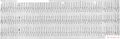
Atrial flutter with 1:1 conduction
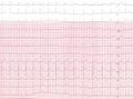
Junctional Ectopic Tachycardia (JEt)
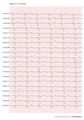
Pacemakerrhythm with retrograde Wenkebach.
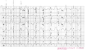
Extreme prolonged QT interval with T wave alternans


Help us!
mail or fax (faxnumber +31-84-755 0017) a typical or difficult ECG and we will add it to the site anonimized.