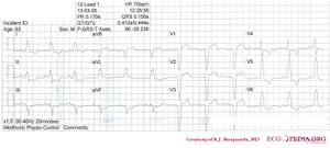
Case 3
Sinusrhythm with left bundle branch block, comparison with an old ECG is mandatory to evaluate whether the LBBB is new (a sign of myocardial infarction) or old.
- Following the 7+2 steps:
- Rhythm
- The ECG shows a regular rhythm with normal P waves (positive in I, III and AVF, negative in AVR), followed by QRS complexes. Sinusrhythm
- Heart rate
- 78 bpm
- Conduction (PQ,QRS,QT)
- PQ: 180ms QRS: 160ms QT: 370ms QTc: 420ms
- Heartaxis
- Negative in III, AVF and AVR, positive QRS complexes in I, II and AVL: horizontal heart axis
- P wave morphology
- The P waves have normal morphology.
- QRS morphology
- Wide QRS complexes with [[[LBBB|left bundle branch block]]] pattern.
- ST morphology
- ST elevation in V1-V3, AVR. ST depression in I, II, III, AVF, V4-6. The Sgarbossa criteria for ischemia in LBBB are not met (nog concordant ST deviation, no ST depression V1-V3, nog discordant ST elevation > 5 mm).
- Compare with the old ECG (not available, so skip this step)
- Conclusion?
- Rhythm
