Answer MI 3

Revision as of 09:15, 11 November 2008 by Drj (talk | contribs) (New page: {{Case| |previouspage= MI 2 |previousname= MI 2 |nextpage=MI 4 |nextname=MI 4 }} '''Where is this myocardial infarction located?''' 700px|thumb|left|ECG MI 3 {{clr}}...)
(diff) ← Older revision | Latest revision (diff) | Newer revision → (diff)
The printable version is no longer supported and may have rendering errors. Please update your browser bookmarks and please use the default browser print function instead.
This page is part of Cases and Examples

Previous ECG: MI 2 | Next ECG: MI 4

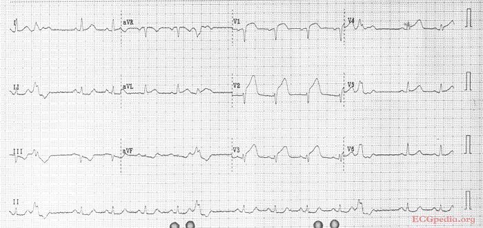
Where is this myocardial infarction located?

ECG MI 3


Answer

Culprit lesion: proximal LAD

  1. sinus rhythm, with 3 ventricular premature beats
  2. about 80 /min
  3. normal conduction
  4. horizontal axis
  5. normal p wave morphology
  6. Q in V1. slow anterior R-wave progression.
  7. ST elevation in V1-V4, AVL. ST depression in II,III,AVF. The ST vector is upright.
  • Conclusion: Anterior MI with proximal LAD occlusion