Answer MI 17
Jump to navigation
Jump to search
| This page is part of Cases and Examples |
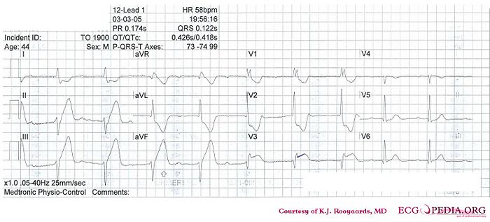
Where is this myocardial infarction located?
Answer
- Following the 7+2 steps:
- Rhythm
- In leads V1 to V3 there is apparantly no relationship between the P waves and the QRS complexes. Furthermore the QRS complexes are broad and have a RBBB pattern (V1:RSR), thus the rhythm is probably a ventricular escape rhythm. Leads V4-V6 however show a narrow complex preced by a P wave, probably the result of a rhythm change to sinus rhythm.
- Hartfrequency
- Use the 'count the squares' method (a bit less than 3 large squares ~> 300-150-100), thus about 58 bpm.
- Conduction (PQ,QRS,QT)
- During sinus beats: PQ-interval=250msec, QRS duration=0.11sec, QT interval=400ms (equals QTc at this rate). The wide complexes have a QRS duration of +- 160 ms.
- Heartaxis
- Negative in AVF and II, thus a left axis deviation.
- P wave morphology
- The p wave is rather difficult to discern. It seems normal in V4-V6 and positive in AVF and negative in AVR.
- QRS morphology
- RBBB pattern in V1 with ST changes (depression precordial leads). Leads V3 and V4 have probably been poled to the right chest. No pathologic Q waves.
- ST morphology
- ST elevation in II,III, and AVF and also in V3 (V3right). Hyperacute T waves with ST depression in V1-V2 an I, AVR and AVL.
- Compare with the old ECG (not available, so skip this step)
- Conclusion?
- Rhythm
Inferior-posterior myocardial infarction with complete AV block and ventricular excape rhythm with RBBB pattern and left axis, followed by sinus-rhythm. Probably RCA occlusion (ST depression in I)
