Answer MI 14: Difference between revisions
Jump to navigation
Jump to search
(New page: ===Answers=== * Describe the ECG according to the 7 + 2 stepplan **Rhythm ***'''This is a regular rhythm and every QRS complex is preceded by a p wave. The p wave is positive in II, III a...) |
No edit summary |
||
| Line 5: | Line 5: | ||
***'''This is a regular rhythm and every QRS complex is preceded by a p wave. The p wave is positive in II, III and AVF and originates form the sinusnode. Conclusion: sinusrhythm.''' | ***'''This is a regular rhythm and every QRS complex is preceded by a p wave. The p wave is positive in II, III and AVF and originates form the sinusnode. Conclusion: sinusrhythm.''' | ||
**Heartfrequency. | **Heartfrequency. | ||
***'''Use the ' | ***'''Use the 'Countingmethod' (6 big grids ~> 300-150-100-75-60-50), so 50/min.''' | ||
** | **Conductiontimes (PQ,QRS,QT) | ||
***'''PQ-tijd=0.16sec (4 | ***'''PQ-tijd=0.16sec (4 small grids), QRS duration=0.10sec, QT time=460ms''' | ||
** | **Heartaxis | ||
***''' | ***'''Positive in I, iso-electric in II, negative in III and AVF. So, a left axis.''' | ||
**P | **P wave morphology | ||
***''' | ***'''The p wave is normal shaped.''' | ||
**QRS | **QRS morphology | ||
***''' | ***'''Conductiondelay right, btu not enough for the RBBB criteria (QRS < 0.12s). Slow R-wave progression in the precordial leads.''' | ||
**ST | **ST morphology | ||
***''' | ***'''ST elevation in II,III and AVF. Reciprocal depression in I, AVR and AVL with negative T waves. Additionally discrete elevation in V2-V5. And ST-elevation in V4R''' | ||
** | **Compare with the old ECG (not available) | ||
** | **conclusion. What is going on? | ||
''' | '''Answer: Inferior wall infarct with right ventricular involvement and: | ||
* | * Sinusbradycardia, probably because the sinusnodebranch, coming form the RCA is lacking perfusion. | ||
* | * Left heart axis | ||
[[ | [[Image:Casus2_2.jpg|thumb|300px|left| the ECG]] | ||
{{clr}} | {{clr}} | ||
[[ | [[Image:Casus2_1.jpg|thumb|300px|left| This is a right lead ECG]] | ||
{{clr}} | {{clr}} | ||
Revision as of 11:26, 6 May 2007
Answers
- Describe the ECG according to the 7 + 2 stepplan
- Rhythm
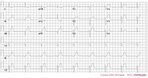
- This is a regular rhythm and every QRS complex is preceded by a p wave. The p wave is positive in II, III and AVF and originates form the sinusnode. Conclusion: sinusrhythm.
- Heartfrequency.
- Use the 'Countingmethod' (6 big grids ~> 300-150-100-75-60-50), so 50/min.
- Conductiontimes (PQ,QRS,QT)
- PQ-tijd=0.16sec (4 small grids), QRS duration=0.10sec, QT time=460ms
- Heartaxis
- Positive in I, iso-electric in II, negative in III and AVF. So, a left axis.
- P wave morphology
- The p wave is normal shaped.
- QRS morphology
- Conductiondelay right, btu not enough for the RBBB criteria (QRS < 0.12s). Slow R-wave progression in the precordial leads.
- ST morphology
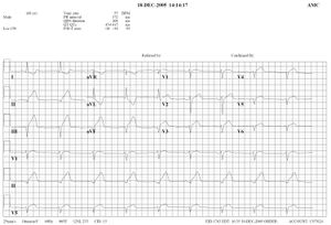
- ST elevation in II,III and AVF. Reciprocal depression in I, AVR and AVL with negative T waves. Additionally discrete elevation in V2-V5. And ST-elevation in V4R
- Compare with the old ECG (not available)
- conclusion. What is going on?
- Rhythm
Answer: Inferior wall infarct with right ventricular involvement and:
- Sinusbradycardia, probably because the sinusnodebranch, coming form the RCA is lacking perfusion.
- Left heart axis

